Comparative Study
. 1997 Jan 15;17(2):834-42.
doi: 10.1523/JNEUROSCI.17-02-00834.1997.

Differential localization of delta glutamate receptors in the rat cerebellum: coexpression with AMPA receptors in parallel fiber-spine synapses and absence from climbing fiber-spine synapses

Affiliations
Comparative Study

Differential localization of delta glutamate receptors in the rat cerebellum: coexpression with AMPA receptors in parallel fiber-spine synapses and absence from climbing fiber-spine synapses

A S Landsend et al. J Neurosci. .

Abstract

The delta 2 glutamate receptors are prominently expressed in Purkinje cells and are thought to play a key role in the induction of cerebellar long-term depression. The synaptic and subsynaptic localization of delta receptors in rat cerebellar cortex was investigated with sensitive and high-resolution immunogold procedures. After postembedding incubation with an antibody raised to a C-terminal peptide of delta 2, high gold particle densities occurred in all parallel fiber synapses with Purkinje cell dendritic spines, whereas other synapses were consistently devoid of labeling. Among the types of immunonegative synapse were climbing fiber synapses with spines and parallel fiber synapses with dendritic stems of interneurons. At the parallel fiber-spine synapse, gold particles signaling delta receptors were restricted to the postsynaptic specialization. By the use of double labeling with two different gold particle sizes, it was shown that delta and AMPA GluR2/3 receptors were colocalized along the entire extent of the postsynaptic specialization without forming separate domains. The distribution of gold particles representing delta receptors was consistent with a cytoplasmic localization of the C terminus and an absence of a significant presynaptic pool of receptor molecules. The present data suggest that the delta 2 receptors are targeted selectively to a subset of Purkinje cell spines and that they are coexpressed with ionotropic receptors in the postsynaptic specialization. This arrangement could allow for a direct interaction between the two classes of receptor.


Figures

Fig. 1.
Fig. 1.
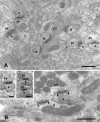
Distribution of δ receptor immunoreactivity at synapses between parallel fibers (Pf) and Purkinje cell spines (s). Seven synapses of this category are shown in the micrograph, and each displays at least three gold particles. The synapse at top center is obliquely cut, the two rows of particles representing receptors exposed at opposite surfaces of the section. Asterisks indicate glial lamellae. Only seven particles are not associated with any postsynaptic density; of these, one is found within a spine (arrow) and two within other intracellular compartments (arrowheads). Inset, Higher magnification of a parallel fiber synapse. Gold particles occur along the entire postsynaptic density (delimited by arrowheads).M, Mitochondrion. Gold particles, 15 nm. Scale bars: 0.5 μm; inset, 0.1 μm.
Fig. 2.
Fig. 2.
Immunolabeling for the δ receptor is absent from synapses between climbing fibers (Cf) and Purkinje cell spines (asterisks) and between mossy fibers (Mf) and granule cell dendritic digits (d). Arrowheads in Bdelimit postsynaptic densities. A and Bare from the same section as Figure 1. B,Insets, Immunolabeling for the δ receptor is abolished after preadsorption with the peptide used for immunization (right; arrows show negative postsynaptic densities) but remains after preadsorption with the peptide used to generate the GluR2/3 antibody (left). Pf, Parallel fibers; s, Purkinje cell spines. Gold particles, 15 nm. Scale bars: A, B, 0.5 μm; insets, 0.25 μm.
Fig. 3.
Fig. 3.
No δ receptor immunolabeling occurs at synapses between parallel fibers (Pf) and dendritic stems (d) or postsynaptic to stellate cell terminals (St), whereas adjacent parallel fiber synapses with spines (s) show dense labeling. Long arrows indicate negative synapses. Arrowhead inA shows gold particle in spine. The stellate cell synapse in B is obliquely cut but can be identified by the presence of flattened vesicles (short arrows).B, Inset, Asymmetric synapse (arrows) established by nerve terminal (T) in stratum oriens in the CA1 of hippocampus. The section was incubated together with the cerebellar section inA. The sizes of gold particles were 15 nm (A and inset in B) or 10 nm (B), the latter giving a slightly higher background labeling than the former. Scale bars: A, B, 0.5 μm;inset, 0.25 μm.
Fig. 4.
Fig. 4.
Tangential (A) and perpendicular (B) distribution of δ receptor immunoreactivity at the parallel fiber–spine synapses. Only synapses with distinct and transversely cut postsynaptic membranes were included in the analysis. Same material as in Figures 1 and 2. A, Immunolabeling occurs along the entire mediolateral extent of the postsynaptic density, but the concentration of particles shows a slight decrease near the margin of the synapse. This decrease can be explained on methodological grounds if one takes into account that the gold particle density at any one point is a function of the antigen concentration within a radius of ∼28 nm (Matsubara et al., 1996). Similarly, the few particles situated lateral to the margin of the postsynaptic thickening can be attributed to epitopes located in the thickening itself. Synapses (n = 68) were selected for analysis only if the radius of the postsynaptic density profile exceeded 150 nm. This should ensure the inclusion of central as well as peripheral parts of the density (the diameter of the postsynaptic density is in the range of 250–500 μm; Palay and Chan-Palay, 1974). The abscissa indicates the mediolateral extent of the postsynaptic density in percentage of distance from the center of the profile (0%) to the margin (100%). B, Gold particles signaling the δ receptor are found predominantly at the postsynaptic side of the postsynaptic membrane (n = 23). The distances between the centers of the gold particles and the midpoint of the postsynaptic plasma membrane were grouped into bins 4 nm wide (bin centers indicated; minus signs denote bins presynaptic to the reference point).
Fig. 5.
Fig. 5.
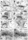
Double labeling with antisera to the δ receptor (30 nm particles in A–F, 5 nm particles inG, H) and GluR2/3 (10 nm inA–F, 15 nm in G,H). Double labeling is found at synapses between parallel fibers (Pf) and Purkinje cell spines (s), whereas climbing fibers (Cf) establish synapses that are single-labeled for GluR2/3 (F). Synapses in G andH are obliquely cut so that parts of the postsynaptic membranes are viewed en face. Some of the 5 nm particles are indicated by arrows. The distance between these particles is typically in the range of 15–30 nm (H,right part). Scale bars: A–F, 0.25 μm;G, H, 0.1 μm.

References

    1. Araki K, Meguro H, Kushiya E, Takayama C, Inoue Y, Mishina M. Selective expression of the glutamate receptor channel delta 2 subunit in cerebellar Purkinje cells. Biochem Biophys Res Commun. 1993;197:1267–1276. - PubMed
    1. Baude A, Nusser Z, Roberts JDB, Mulvihill E, McIlhinney RAJ, Somogyi P. The metabotropic glutamate receptor (mGluR1α) is concentrated at perisynaptic membrane of neuronal subpopulations as detected by immunogold reaction. Neuron. 1993;11:771–787. - PubMed
    1. Baude A, Nusser Z, Molnár E, McIlhinney RAJ, Somogyi P. High-resolution immunogold localization of AMPA-type glutamate receptor subunits at synaptic and nonsynaptic sites in the rat hippocampus. Neuroscience. 1995;69:1031–1055. - PubMed
    1. Bennet JA, Dingledine R. Topology profile for a glutamate receptor: three transmembrane domains and a channel-lining reentrant membrane loop. Neuron. 1995;14:373–384. - PubMed
    1. Chaudhry FA, Lehre KP, van Lookeren Campagne M, Ottersen OP, Danbolt NC, Storm-Mathisen J. Glutamate transporters in glial plasma membranes: highly differentiated localizations revealed by quantitative ultrastructural immunocytochemistry. Neuron. 1995;15:711–720. - PubMed

Publication types

LinkOut - more resources