. 1996 Dec 15;16(24):7812-20.
doi: 10.1523/JNEUROSCI.16-24-07812.1996.

Translocation of RNA granules in living neurons

Affiliations

Translocation of RNA granules in living neurons

R B Knowles et al. J Neurosci. .

Abstract

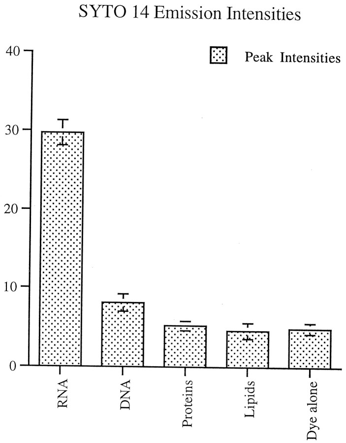
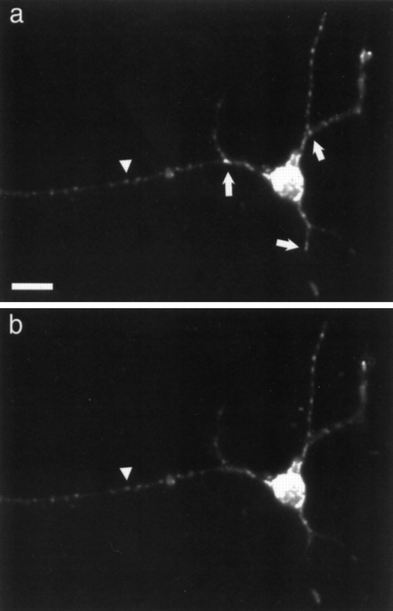
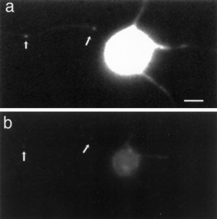
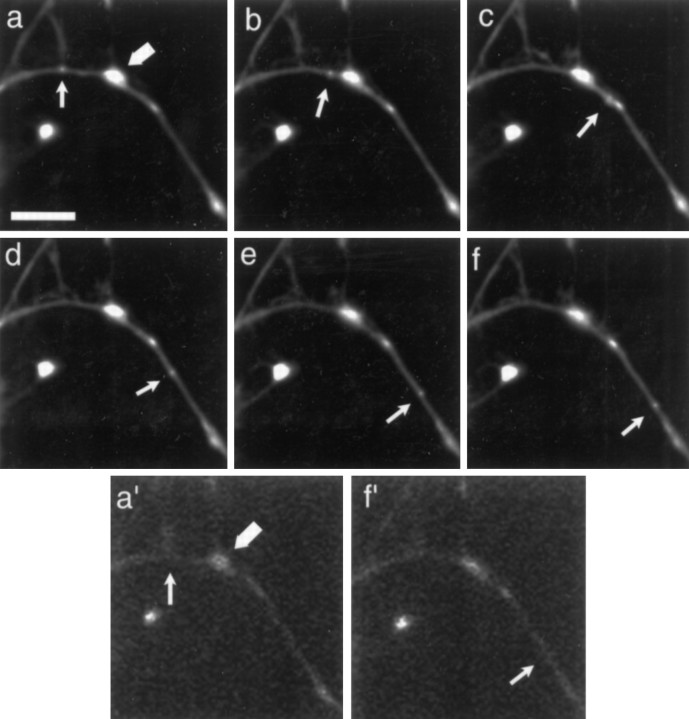
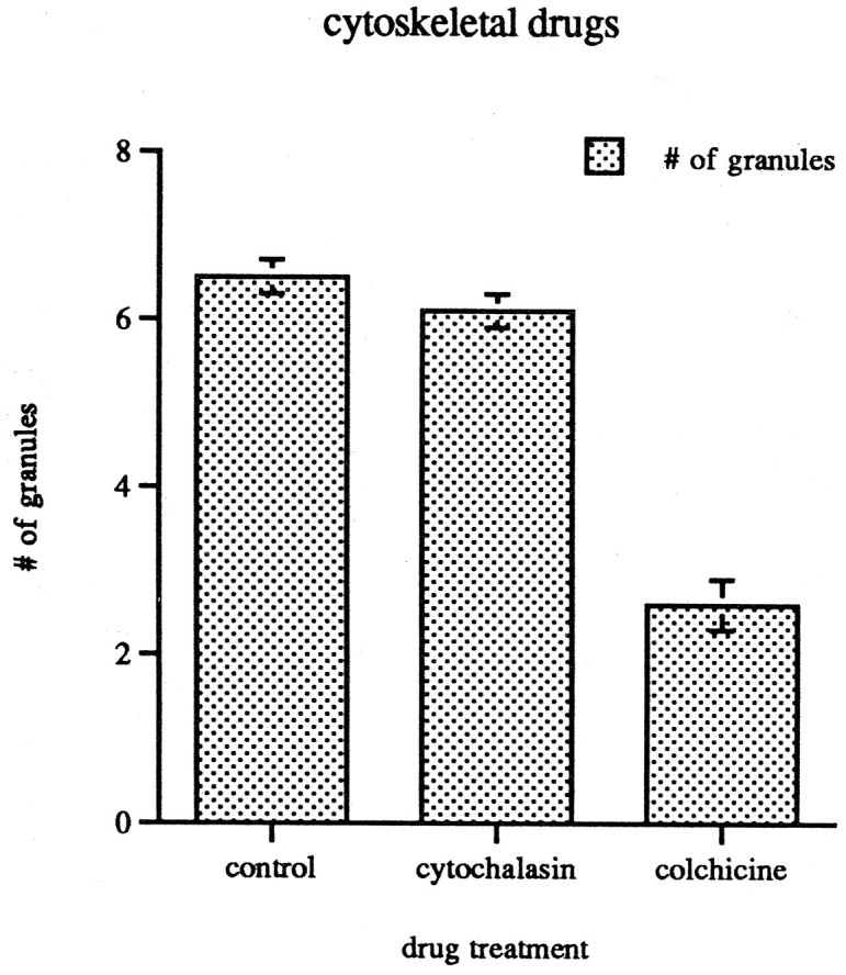
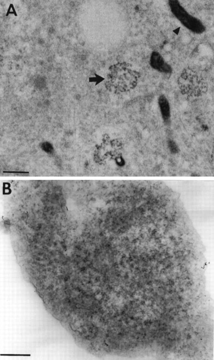
Sorting of RNAs to specific subcellular loci occurs in diverse settings from fly oocytes to mammalian neurons. Using the membrane-permeable nucleic acid stain SYTO 14, we directly visualized the translocation of endogenous RNA in living cells. Labeled RNA was distributed nonrandomly as discrete granules in neuronal processes. The labeled granules colocalized with poly(A+) mRNA, with the 60S ribosomal subunit, and with elongation factor 1alpha, suggesting that granules represent a translational unit. A subset of labeled granules colocalized with beta-actin mRNA. Correlative light and electron microscopy indicated that the fluorescent granules corresponded to clusters of ribosomes at the ultrastructural level. Poststaining of sections with heavy metals confirmed the presence of ribosomes within these granules. In living neurons, a subpopulation of RNA granules was motile during the observation period. They moved at an average rate of 0.1 microm/sec. In young cultures their movements were exclusively anterograde, but after 7 d in culture, one-half of the motile granules moved in the retrograde direction. Granules in neurites were delocalized after treatment with microtubule-disrupting drugs. These results raise the possibility of a cellular trafficking system for the targeting of RNA in neurons.


Figures

Fig. 1.
Fig. 1.
Emission intensity profiles of RNA, DNA, proteins, and lipids incubated with SYTO 14 and scanned at an absorbance of 500 nm. Peak emission intensity for RNA was sixfold greater than that for either proteins or lipids. It differed from proteins, lipids, and DNA with a significance of p < 0.001. The emission intensity for DNA differed from proteins and lipids with a significance of p < 0.05.
Fig. 2.
Fig. 2.
Cortical neurons colabeled with SYTO 14 (a) and Mitotracker (b).Arrowhead indicates mitochondria that colocalize with both markers. Small arrows indicate granules only observed with SYTO 14 labeling. Scale bar, 20 μm.
Fig. 3.
Fig. 3.
Cortical neurons labeled with SYTO 14 before (a) and after (b) RNase treatment.Arrows indicate SYTO 14-labeled granules. Scale bar, 10 μm.
Fig. 4.
Fig. 4.
Cortical neurons labeled with SYTO 14 and translational components. a, SYTO 14-labeled granules (small arrows) and mitochondria (large arrow) staining in neurites. b, Poly(A+) mRNA-containing granules detected in neurites (small arrows) and no signal at location of mitochondria (large arrow). Scale bar, 5 μm. c, SYTO 14-labeled granules in neurites. d, β-Actin mRNA-containing granules in neurites. Examples of colocalization are indicated with arrows. Scale bar, 5 μm.e, SYTO 14-labeled granule staining in neurites.f, Ribosomal 60S subunit labeling in neurites. Examples of colocalization are indicated with arrows.Arrowhead in e points to start of axon-like neurite. Scale bar, 10 μm. g, SYTO 14-labeled granule staining in neurites. h, EF1α labeling in neurites. Examples of colocalization are indicated witharrows. Phase contrast showed that indicated granules were located on neurite branches (data not shown). Scale bar, 5 μm.
Fig. 5.
Fig. 5.
Time-lapse analysis of SYTO 14-labeled small granule movement in neurites. a–f, Images after granule (small arrow) and mitochondria (large arrow) at times 0, 1, 2, 3, 4, 5 min. a′, Image taken of mitochondria (large arrow) just before time-lapse. f′, Image taken of mitochondria just after time-lapse. Scale bar, 10 μm.
Fig. 6.
Fig. 6.
Distribution of SYTO 14-labeled granules in 40 μm segments in the neurite shafts of 4-d-old cortical neuronal cultures. Treatment with cytochalasin-D does not significantly alter the distribution of RNA granules, whereas treatment with colchicine decreases the number by 62% (p < 0.001).
Fig. 7.
Fig. 7.
A, Cortical neurons labeled with SYTO 14, photoconverted in the presence of DAB, and viewed with an electron microscope. Arrows point to electron-dense DAB reaction product that corresponds to the precise location of the SYTO 14 dye. The labeled structures appear as clusters of ribosomes (arrow) and organelles that seem to be mitochondria (arrowhead). Sections are 90 μm thick. Scale bar, 500 nm. B, Electron micrograph of a single RNA granule. The site shown here precisely corresponds to a SYTO 14-labeled granule in a culture of cortical neurons. Scale bar, 100 nm.

Similar articles

Cited by

References

    1. Ainger K, Avossa D, Morgan F, Hill SJ, Barry C, Barbarese E, Carson JH. Transport and localization of exogenous MBP mRNA microinjected into oligodendrocytes. J Cell Biol. 1993;123:431–441. - PMC - PubMed
    1. Atkinson S, Doberstein SK, Pollard TD. Moving off the beaten track. Curr Biol. 1992;2:326–328. - PubMed
    1. Baas PW, Black MW, Banker GA. Changes in microtubule polarity orientation during the development of hippocampal neurons in culture. J Cell Biol. 1989;109:3085–3094. - PMC - PubMed
    1. Banker GA, Waxman AB. Hippocampal neurons generate natural shapes in cell culture. In: Lasek RJ, Black MM, editors. Studies of the intrinsic determinants of neuronal form and function. Liss; New York: 1988. pp. 61–82.
    1. Barbarese E, Koppel DE, Deutscher MP, Smith CL, Ainger K, Morgan F, Carson JH. Protein translation components are colocalized in granules in oligodendrocytes. J Cell Sci. 1995;108:2781–2790. - PubMed

Publication types

LinkOut - more resources