Polygenic disease and retinitis pigmentosa: albinism exacerbates photoreceptor degeneration induced by the expression of a mutant opsin in transgenic mice
- PMID: 8987813
- PMCID: PMC6579236
- DOI: 10.1523/JNEUROSCI.16-24-07853.1996
Polygenic disease and retinitis pigmentosa: albinism exacerbates photoreceptor degeneration induced by the expression of a mutant opsin in transgenic mice
Abstract
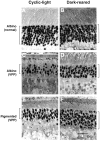
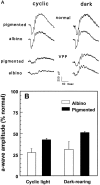
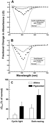
Expression of a mouse opsin transgene containing three point mutations (V20G, P23H, and P27L; termed VPP) causes a progressive photoreceptor degeneration that resembles in many important respects that seen in patients with autosomal dominant retinitis pigmentosa caused by a P23H point mutation. We have attempted to determine whether the degree of degeneration induced by expression of the transgene is influenced by albinism, a genetically mediated recessive trait that results in a deficiency in melanin formation in pigmented tissues throughout the body. Litters of albino and pigmented mice (normal as well as transgenic) were reared in either darkness or cyclic light. Retinal structure and function were evaluated by light microscopy, electroretinography (ERG), and retinal densitometry. The data were consistent in demonstrating that at similar ages, the extent of photoreceptor degeneration was greater in transgenic albino animals than in their pigmented counterparts. The albino VPP mice had significantly fewer cell bodies in the outer nuclear layer of the retina, a larger reduction in ERG amplitude, and a lower rhodopsin content in the rod photoreceptors. These structural and functional differences could not be attributed to the greater level of retinal illumination experienced by the albino retina under normal ambient conditions, because they persisted when pigmented and albino mice were reared in darkness from birth. Although the explanation remains unclear, our findings indicate that the rate of photoreceptor degeneration in VPP mice is adversely affected by the existence of the albino phenotype, a factor that may have implications for the counseling of human patients with retinitis pigmentosa and a familial history of other genetic disorders.
Figures


Similar articles
-
Light-induced acceleration of photoreceptor degeneration in transgenic mice expressing mutant rhodopsin.Invest Ophthalmol Vis Sci. 1996 Apr;37(5):775-82. Invest Ophthalmol Vis Sci. 1996. PMID: 8603862
-
Simulation of human autosomal dominant retinitis pigmentosa in transgenic mice expressing a mutated murine opsin gene.Proc Natl Acad Sci U S A. 1993 Jun 15;90(12):5499-503. doi: 10.1073/pnas.90.12.5499. Proc Natl Acad Sci U S A. 1993. PMID: 8516292 Free PMC article.
-
Functional abnormalities in transgenic mice expressing a mutant rhodopsin gene.Invest Ophthalmol Vis Sci. 1995 Jan;36(1):62-71. Invest Ophthalmol Vis Sci. 1995. PMID: 7822160
-
Transgenic mice with a rhodopsin mutation (Pro23His): a mouse model of autosomal dominant retinitis pigmentosa.Neuron. 1992 Nov;9(5):815-30. doi: 10.1016/0896-6273(92)90236-7. Neuron. 1992. PMID: 1418997
-
Expression of a mutant opsin gene increases the susceptibility of the retina to light damage.Vis Neurosci. 1997 Jan-Feb;14(1):55-62. doi: 10.1017/s0952523800008750. Vis Neurosci. 1997. PMID: 9057268
Cited by
-
The effect of peripherin/rds haploinsufficiency on rod and cone photoreceptors.J Neurosci. 1997 Nov 1;17(21):8118-28. doi: 10.1523/JNEUROSCI.17-21-08118.1997. J Neurosci. 1997. PMID: 9334387 Free PMC article.
-
Longitudinal fundus and retinal studies with SD-OCT: a comparison of five mouse inbred strains.Mamm Genome. 2013 Jun;24(5-6):198-205. doi: 10.1007/s00335-013-9457-z. Epub 2013 May 17. Mamm Genome. 2013. PMID: 23681115
-
Electrophysiological analysis of visual function in mutant mice.Doc Ophthalmol. 2003 Jul;107(1):13-36. doi: 10.1023/a:1024448314608. Doc Ophthalmol. 2003. PMID: 12906119 Review.
-
Retinal degeneration in two lines of transgenic S334ter rats.Exp Eye Res. 2011 Mar;92(3):227-37. doi: 10.1016/j.exer.2010.12.001. Epub 2010 Dec 11. Exp Eye Res. 2011. PMID: 21147100 Free PMC article.
-
Progressive Retinal Degeneration Increases Cortical Response Latency of Light Stimulation but Not of Electric Stimulation.Transl Vis Sci Technol. 2022 Apr 1;11(4):19. doi: 10.1167/tvst.11.4.19. Transl Vis Sci Technol. 2022. PMID: 35446408 Free PMC article.
References
-
- Al-Ubaidi MR, Pittler SJ, Champagne MS, Triantafyllos JT, McGinnis JF, Baehr W. Mouse opsin: gene structure and molecular basis of multiple transcripts. J Biol Chem. 1990;265:20563–20569. - PubMed
-
- Applebury ML. Variations in retinal degeneration. Curr Biol. 1992;2:113–115. - PubMed
-
- Baehr W, Falk JD, Bugra K, Triantafyllos JT, McGinnis JF. Isolation and analysis of the mouse opsin gene. FEBS Lett. 1988;238:253–256. - PubMed
-
- Berson EL. Retinitis pigmentosa. Invest Ophthalmol Vis Sci. 1993;34:1659–1676. - PubMed
-
- Berson EL, Rosner B, Sandberg MA, Dryja TP. Ocular findings in patients with autosomal dominant retinitis pigmentosa and a rhodopsin gene defect (pro-23-his). Arch Ophthalmol. 1991;109:92–101. - PubMed
Publication types
MeSH terms
Substances
Grants and funding
LinkOut - more resources
Full Text Sources
