. 1996 Dec 15;16(24):7892-901.
doi: 10.1523/JNEUROSCI.16-24-07892.1996.

Neuronal nicotinic receptor expression in sensory neurons of the rat trigeminal ganglion: demonstration of alpha3beta4, a novel subtype in the mammalian nervous system

Affiliations

Neuronal nicotinic receptor expression in sensory neurons of the rat trigeminal ganglion: demonstration of alpha3beta4, a novel subtype in the mammalian nervous system

C M Flores et al. J Neurosci. .

Abstract

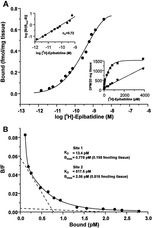
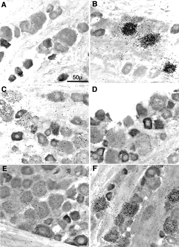
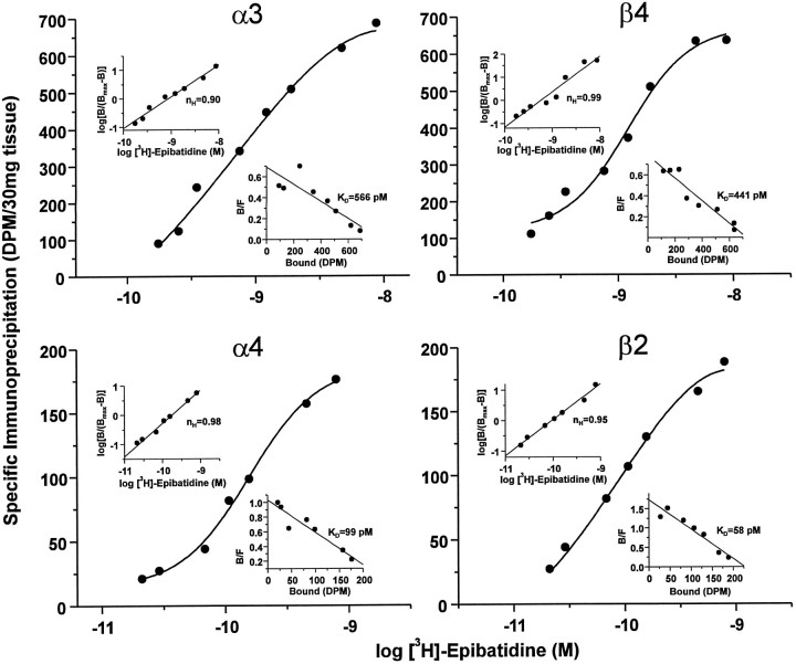
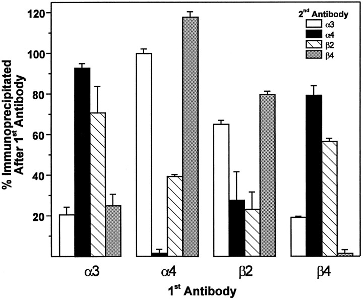
The identification of a family of neuronal nicotinic receptor subunit genes establishes the potential for multiple subtypes with diverse physiological functions. Virtually all of the high affinity nicotinic receptors measured to date in the rodent CNS are composed of alpha4 and beta2 subunits only. However, the demonstration of other subunit transcripts in a variety of central and peripheral nervous tissues suggests a greater degree of receptor subtype heterogeneity than so far has been elucidated. The purpose of the present studies was to determine at the mRNA and protein levels which neuronal nicotinic receptor subunits are expressed by sensory neurons of the rat trigeminal ganglion and in what combinations these gene products associate to form neuronal nicotinic receptor subtypes in this tissue. Radioreceptor binding analysis indicated that in the adult rat trigeminal ganglion there exist at least two nicotinic receptor binding sites with differing affinities for [3H]-epibatidine. In situ hybridization histochemical studies revealed the existence of mRNA encoding the alpha3, alpha4, alpha5, beta2, and beta4 subunits, but not the alpha2 subunit. Immunoprecipitation with subunit-specific antisera demonstrated that each of the subunits present in the ganglion at the mRNA level is a constituent of nicotinic receptors capable of binding 3H-epibatidine. Various applications of these approaches yielded strong evidence that, in addition to alpha4beta2, which is thought to be the predominant neuronal nicotinic receptor subtype in the rodent CNS, trigeminal sensory neurons express as the principal subtype alpha3beta4, which has not been demonstrated previously in mammalian nervous tissue.


Figures

Fig. 1.
Fig. 1.
Neuronal nicotinic receptor binding sites in the rat trigeminal ganglion. Saturation binding analysis of neuronal nicotinic receptors labeled with [3H]-epibatidine in membrane homogenates of rat trigeminal ganglion. Aliquots of homogenized, washed trigeminal ganglion membranes equivalent to 20 mg of original tissue weight were incubated in triplicate with [3H]-epibatidine at the indicated concentrations (1.4 pm to 3.9 nm) in the absence or presence of 300 μm nicotine bitartrate (to define nonspecific binding). The median value of each triplicate measurement was used to generate all data. A, Semilog plot of the data expressed as bound receptor (fmol/mg tissue) and calculated as the difference between total and nonspecific binding. Top left inset, Hill plot, including the calculated Hill coefficient (nH), of the transformed data. Bottom right inset, Linear plot of the specific (squares) and nonspecific (circles) binding at each concentration of [3H]-epibatidine.B, Rosenthal plot of the data shown in A. Data were analyzed by nonlinear regression with LIGAND (Munson and Rodbard, 1980) and were best fit to a two-site model (ANOVA,F1,8 = 51.9; p < 0.001), as indicated by the dashed lines. The calculated receptor density (Bmax) and equilibrium dissociation constant (KD) values for each site are indicated. Data are representative of an experiment performed four times.
Fig. 2.
Fig. 2.
Neuronal nicotinic receptor subunit mRNA expression in sensory neurons of the rat trigeminal ganglion. Combinedin situ hybridization histochemistry and immunocytochemistry for neuronal nicotinic receptor subunit mRNAs and peripherin in the rat trigeminal ganglion. Bright-field images are of 20-μm-thick frozen sections of adult male rat trigeminal ganglion sequentially processed for in situ hybridization by a35S-labeled riboprobe complimentary to the mRNA encoding the α2 (A), α3 (B), α4 (C), α5 (D), β2 (E), or β4 (F) neuronal nicotinic receptor subunit (represented by black grains), followed by immunocytochemistry with an anti-peripherin rabbit serum (seen asdarkly stained cells). After ABC (peroxidase) color development, slides were subjected to emulsion autoradiography, counterstained, coverslipped, and photographed. Total magnification, 40×; scale is indicated in A. Photographs are representative of experiments performed at least five times for each subunit transcript with two separate and nonoverlapping probes.
Fig. 3.
Fig. 3.
Subunit constituents of neuronal nicotinic receptors in the rat trigeminal ganglion. Immunoprecipitation of neuronal nicotinic receptors from rat trigeminal ganglion. Aliquots of [3H]-epibatidine-labeled, Triton X-100-solubilized rat trigeminal ganglion membranes equivalent to 30 mg of original tissue weight were incubated with rabbit antisera specific for each of the neuronal nicotinic receptor subunits indicated or normal rabbit serum (NRS) and precipitated with Pansorbin cells by centrifugation. Specific immunoprecipitation was calculated to be the difference between that obtained with each subunit-specific serum and that obtained with NRS. Data from three to seven experiments are expressed as the mean ± SEM in dpm/30 mg tissue.
Fig. 4.
Fig. 4.
Binding parameters of neuronal nicotinic receptor subtypes in the rat trigeminal ganglion. Saturation binding analysis of neuronal nicotinic receptors labeled with [3H]-epibatidine and immunoprecipitated with subunit-specific rabbit antisera. Aliquots of Triton X-100-solubilized rat trigeminal ganglion membranes equivalent to 30 mg of original tissue weight were labeled with [3H]-epibatidine at the indicated concentrations (0.175–8.7 nm forα3 and β4; 29–792 pm forα4 and β2), incubated with rabbit antisera specific for each of the neuronal nicotinic receptor subunits indicated or normal rabbit serum (NRS), and then precipitated with Pansorbin cells by centrifugation. Specific immunoprecipitation was calculated to be the difference between that obtained with each subunit-specific serum and that obtained with NRS at each concentration of [3H]-epibatidine. Each quadrant depicts a semilog plot of the data for each of the four antisera tested, expressed as specific immunoprecipitation in dpm/30 mg of tissue. Top left insets, Hill plot, including the calculated Hill coefficient (nH), of the transformed data. Bottom right insets, Rosenthal plots, including calculated equilibrium dissociation constant (KD) values, of the transformed data. Data were analyzed by nonlinear regression with LIGAND (Munson and Rodbard, 1980) and, for each subunit, were best fit to a one-site model as indicated by the single solid lines (circles). Because of the tremendous amounts of tissue, radioactivity, and antibody required, this experiment was performed only once.
Fig. 5.
Fig. 5.
Subunit composition of neuronal nicotinic receptor subtypes in the rat trigeminal ganglion. Double immunoprecipitation of neuronal nicotinic receptors from rat trigeminal ganglion. Aliquots of [3H]-epibatidine-labeled, Triton X-100-solubilized rat trigeminal ganglion membranes equivalent to 30 mg of original tissue weight were incubated with rabbit antisera specific for each of the neuronal nicotinic receptor subunits (indicated on theabscissa as 1st Antibody) or normal rabbit serum (NRS) and precipitated with Pansorbin cells by centrifugation. The resulting supernatant was reprecipitated in triplicate with a second antibody (indicated by theinset as 2nd Antibody). Data (mean ± SEM) are expressed as the percentage immunoprecipitated after the first antibody, which was calculated as the ratio of the amount of specific immunoprecipitation obtained by the second antibody after preclearing with the first antibody divided by the amount of specific immunoprecipitation obtained by the second antibody after preclearing with NRS (see text for rationale and interpretation). Data are representative of an experiment performed four times.
Fig. 6.
Fig. 6.
Colocalization of α3 and β4 mRNA in individual neurons of the rat trigeminal ganglion. Double isotopic and nonisotopicin situ hybridization histochemistry for α3 and β4 neuronal nicotinic receptor subunit mRNAs in the rat trigeminal ganglion. Bright-field (A) and epi-polarized (B) images are of the same 20-μm-thick frozen section of adult male rat trigeminal ganglion simultaneously hybridized with a 35S-labeled riboprobe complimentary to the mRNA encoding the α3 subunit (represented by black grains in A and white grains inB) and a biotinylated riboprobe complimentary to the β4 subunit (seen as darkly stained cells inA). After ABC (alkaline phosphatase) color development, slides were subjected to emulsion autoradiography, coverslipped, and photographed. Total magnification, 40×; scale is indicated inB. Photographs are representative of an experiment performed three times.

References

    1. Anand R, Conroy WG, Schoepfer R, Whiting P, Lindstrom J. Neuronal nicotinic acetylcholine receptors expressed in Xenopus oocytes have a pentameric quaternary structure. J Biol Chem. 1991;266:11192–11198. - PubMed
    1. Angelotti TP, Macdonald RL. Assembly of GABAA receptor subunits: α1β1 and α1β1δ2s subunits produce unique ion channels with dissimilar single-channel properties. J Neurosci. 1993;13:1429–1440. - PMC - PubMed
    1. Baccaglini PI, Cooper E. Influences on the expression of acetylcholine receptors on rat nodose neurones in culture. J Physiol (Lond) 1982;324:441–451. - PMC - PubMed
    1. Boulter J, Evans K, Goldman D, Martin G, Treco D, Heinemann S, Patrick J. Isolation of a cDNA clone coding for a possible neural nicotinic acetylcholine receptor α-subunit. Nature. 1986;319:368–374. - PubMed
    1. Boulter J, Connolly JG, Deneris ES, Goldman D, Heinemann S, Patrick J. Functional expression of two neuronal nicotinic acetylcholine receptors from cDNA clones identifies a gene family. Proc Natl Acad Sci USA. 1987;84:7763–7767. - PMC - PubMed

Publication types

LinkOut - more resources