. 1996 Dec 15;16(24):7910-9.
doi: 10.1523/JNEUROSCI.16-24-07910.1996.

Matrix metalloproteinase-9 (MMP-9) is synthesized in neurons of the human hippocampus and is capable of degrading the amyloid-beta peptide (1-40)

Affiliations

Matrix metalloproteinase-9 (MMP-9) is synthesized in neurons of the human hippocampus and is capable of degrading the amyloid-beta peptide (1-40)

J R Backstrom et al. J Neurosci. .

Abstract

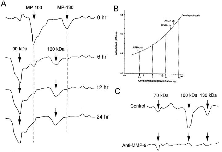
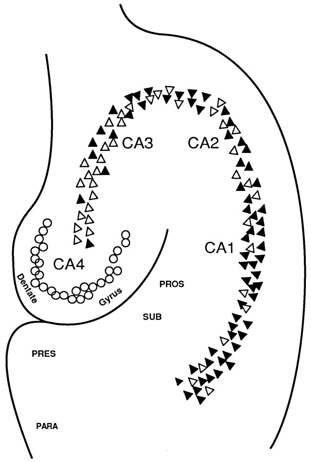
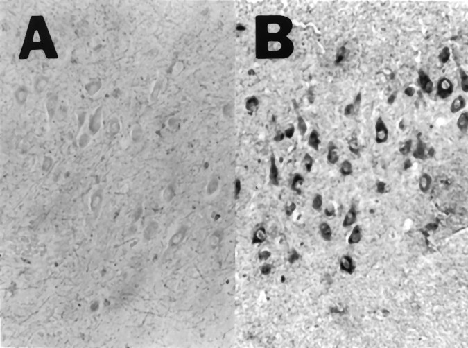
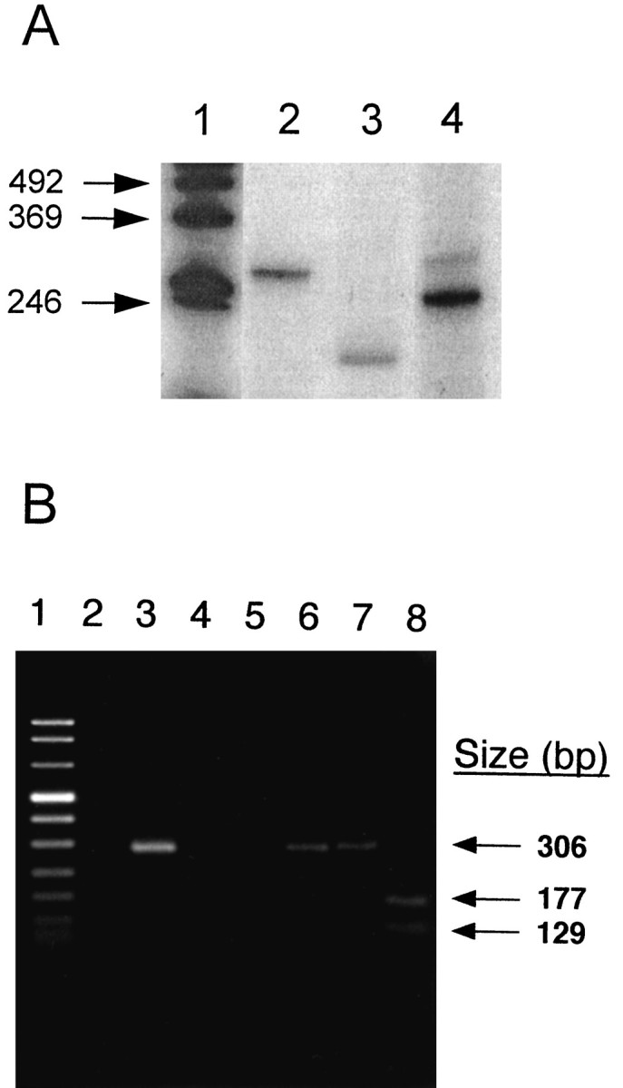
We reported earlier that the levels of Ca2+-dependent metalloproteinases are increased in Alzheimer's disease (AD) specimens, relative to control specimens. Here we show that these enzymes are forms of the matrix metalloproteinase MMP-9 (EC3.4.24. 35) and are expressed in the human hippocampus. Affinity-purified antibodies to MMP-9 labeled pyramidal neurons, but not granular neurons or glial cells. MMP-9 mRNA is expressed in pyramidal neurons, as determined with digoxigenin-labeled MMP-9 riboprobes, and the presence of this mRNA is confirmed with reverse transcriptase PCR. The cellular distribution of MMP-9 is altered in AD because 76% of the total 100 kDa enzyme activity is found in the soluble fraction of control specimens, whereas only 51% is detectable in the same fraction from AD specimens. The accumulated 100 kDa enzyme from AD brain is latent and can be converted to an active form with aminophenylmercuric acetate. MMP-9 also is detected in close proximity to extracellular amyloid plaques. Because a major constituent of plaques is the 4 kDa beta-amyloid peptide, synthetic Abeta1-40 was incubated with activated MMP-9. The enzyme cleaves the peptide at several sites, predominantly at Leu34-Met35 within the membrane-spanning domain. These results establish that neurons have the capacity to synthesize MMP-9, which, on activation, may degrade extracellular substrates such as beta-amyloid. Because the latent form of MMP-9 accumulates in AD brain, it is hypothesized that the lack of enzyme activation contributes to the accumulation of insoluble beta-amyloid peptides in plaques.


Figures

Fig. 1.
Fig. 1.
Activation of latent MMP-9 from the hippocampus.A, Densitometric scan of a substrate gel with gelatin as the substrate. Aliquots of the inhibitor-treated soluble brain fraction were incubated in the absence (−) or presence (+) of 1 mmAPMA for 0, 6, 12, and 24 hr at 37°C. B, Gelatinase activity of a soluble brain fraction with biotinylated gelatin as the substrate in a plate assay. Aliquots of the inhibitor-treated sample were preincubated with 1 mm APMA for 1 or 3 hr at 37°C (arrows). The control for endogenous metalloproteinase activity included a sample incubated in the absence of APMA (0 hr, arrow). Chymotryptic activities were determined in the plate assay with the indicated nanogram amounts of α-chymotrypsin. The experiments were performed three times in triplicate with a 10% SEM. See Materials and Methods for the calculation of specific activities. C, Immunodepletion of enzyme activities with a specific monoclonal antibody to the latent form of MMP-9 (Ab-2, Oncogene Science). Densitometric scans of substrate electrophoretic gels with samples incubated with or without specific monoclonal antibodies to MMP-9. (All experiments were performed on specimens from three AD patients, and samples from patient 206 were used for the illustration.)
Fig. 2.
Fig. 2.
Localization of immunoreactive MMP-9 in the human hippocampus. A, Control section demonstrating that reactivity was below the level of detection (200×). B, AD section illustrating reactivity in pyramidal cells (200×). Glial cells and perivascular areas were unstained. C, Higher magnification of pyramidal neuron from AD section showing granular accumulation of immunoreactive MMP-9 in the cytoplasm.D, Pyramidal neuron showing stained material extending into the neurite. E, Senile plaque illustrating positively stained cellular process (arrowheads).F, Neuritic processes labeled with Bielschowsky stain. (Samples from AD patients 107, 342, 538, 595, and 602 and from control patients 95, 559, and 612 were investigated. Specimens from 595 and 559 are used for illustration.)
Fig. 3.
Fig. 3.
Summary of anti-MMP-9 staining in the human hippocampus. Pyramidal neurons that were positively stained with anti-MMP-9 (filled triangles) were found in the CA1–CA3 subfields from AD sections. Increasing numbers of unstained neurons (open triangles) were seen from the CA1–CA3 regions. Neurons in the CA4 subfield, as well as granule neurons (open circles) in the dentate gyrus, were unstained.PRES, Presubiculum; PROS, prosubiculum;SUB, subiculum; PARA, parahippocampal gyrus. (Specimens from AD patients 107, 595, and 602 were used for the studies.)
Fig. 4.
Fig. 4.
The antisense MMP-9 riboprobe labels pyramidal neurons in the human hippocampus. The Alzheimer sections were treated with sense (A) or antisense (B) riboprobes. The CA3 region of the hippocampus is illustrated. (Specimens from patient 107 were used for the illustration.)
Fig. 5.
Fig. 5.
Reverse transcriptase-PCR of hippocampus RNA.A, PCR amplification of the MMP-9 active site from an AD sample (No. 206). Lane 1, Size standards; lanes 2, 4, 275 bp fragment of the human thymidylate synthase gene and 252 bp fragment of the human β-actin gene, used as control amplifications, respectively. Lane 3, 211 bp fragment of the MMP-9 active site. B, PCR amplification of the 5′ region of MMP-9 from a normal human cDNA pool. Lane 1, Size standards; lane 2, amplification of the hippocampus cDNA. The remaining portion of the sample was electrophoresed in a separate gel, and the 306 bp DNA was removed, purified, and PCR-amplified. Lane 3 illustrates the reamplification of this 306 bp product. Lanes 4,5, Water controls for the first and second PCR amplifications, respectively. Lane 6, Gel-purified PCR fragment; lanes 7, 8, the gel-purified fragment treated with ApaI (negative control) andPvuII, respectively. The arrows indicate the positions of the 306 bp fragment and the 177 and 129 bp digestion fragments from PvuII.
Fig. 6.
Fig. 6.
Summary of the results from the digestion of Aβ1–40 by MMP-9. Reverse-phase HPLC was used to separate the peptides, and the sequences of the digestion products were determined by mass spectroscopy and amino acid sequencing. The major cleavage site (arrow above line, Leu34-Met35) and minor cleavage sites (arrows below line, Lys16-Leu17, Ala30-Ile31, and Gly37-Gly38) are indicated. Theboxed amino acids represent the region of Aβ within the membrane. The m/z ratios were 4328.9 (Aβ1–40), 3786.2 (Aβ1–34), 560.7 (Aβ35–40), 3390.6 (Aβ1–30), and 1954.5 (Aβ1–16).

References

    1. Abraham CR, Selkoe DJ, Potter H. Immunochemical identification of the serine proteinase inhibitor, alpha-1-antichymotrypsin, in the brain amyloid deposit of Alzheimer’s disease. Cell. 1988;52:487–501. - PubMed
    1. Aisen PS, Davis KL. Inflammatory mechanisms in Alzheimer’s disease: implications for therapy. Am J Psychiatry. 1994;151:1105–1113. - PubMed
    1. Backstrom JR, Tökés ZA. The 84 kDa form of human matrix metalloproteinase-9 (MMP-9) degrades substance P and gelatin. J Neurochem. 1995;64:1312–1318. - PubMed
    1. Backstrom JR, Miller CA, Tökés ZA. Characterization of neutral proteinases from Alzheimer-affected and control brain specimens: identification of calcium-dependent metalloproteinases from the hippocampus. J Neurochem. 1992;58:983–992. - PubMed
    1. Breitner JCS, Gau BA, Welsh KA, Plassman BL, McDonald WM, Helms MJ, Anthony JC. Inverse association of anti-inflammatory treatments and Alzheimer’s disease: initial results of a co-twin control study. Neurology. 1994;44:227–232. - PubMed

Publication types

MeSH terms