. 1996 Dec 15;16(24):8019-26.
doi: 10.1523/JNEUROSCI.16-24-08019.1996.

Influence of cocaine on the JAK-STAT pathway in the mesolimbic dopamine system

Affiliations

Influence of cocaine on the JAK-STAT pathway in the mesolimbic dopamine system

M T Berhow et al. J Neurosci. .

Abstract

Chronic exposure to cocaine produces characteristic biochemical adaptations within the rat ventral tegmental area (VTA), a brain region rich in dopaminergic neurons implicated in the reinforcing and locomotor-activating properties of cocaine. Some of these changes are mimicked by chronic ciliary neurotrophic factor (CNTF) infusions into the same brain area. We show in this study that chronic cocaine treatment regulates the signal transduction pathway used by CNTF specifically in the VTA. There is an increase in immunoreactivity of Janus kinase (JAK2), a CNTF-regulated protein tyrosine kinase, in the VTA after chronic but not acute cocaine administration. This increase is not seen in the nearby substantia nigra or several other brain regions studied. Furthermore, this increase in JAK2 is not seen after chronic administration of other psychotropic drugs and was not observed for JAK1. The increase in JAK2 levels is associated with an increased responsiveness of the system to acute CNTF infusion into the VTA, as measured by induction in this brain region of signal transducers and activators of transcription (STAT) DNA binding activity and of Fos-like proteins, two known functional endpoints of JAK activation. Double-labeling immunohistochemical studies show that JAK2 immunoreactivity in the VTA is enriched in dopaminergic and nondopaminergic cells, both of which exhibit increased JAK2 immunoreactivity after chronic cocaine treatment. These findings suggest a scheme whereby some of the effects of chronic cocaine on VTA dopaminergic neurons are mediated directly by regulation of the JAK-STAT pathway in these cells, as well as perhaps indirectly by regulation of this pathway in nondopaminergic cells.


Figures
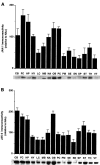
Fig. 1.
Fig. 1.
Regional distribution of (A) JAK1 and (B) JAK2 immunoreactivity in rat brain. Aliquots (20 μg of protein) of SDS-solubilized extracts were subjected to SDS-PAGE, and resulting gels were processed for blot immunolabeling of JAK1 or JAK2 using anti-JAK1 or anti-JAK2 antibodies as described in Materials and Methods. The top panels ofA and B summarize the data (mean ± SEM) obtained from four animals. All data are expressed as percentage of immunoreactivity relative to nucleus accumbens (NAc) normalized to 100%. CB, Cerebellum; FC, frontal cortex; HP, hippocampus; HY, hypothalamus; LC, locus coeruleus; MB, midbrain; NA, nucleus accumbens; OB, olfactory bulb; PC, parietal cortex; PM, pons medulla; SE, septum; SN, substantia nigra; SP, spinal cord; ST, striatum;TH, thalamus; VT, ventral tegmental area. Each panel shows portions of resulting immunoblots obtained from a representative rat.
Fig. 2.
Fig. 2.
Regulation of JAK2 immunoreactivity by chronic cocaine. A, Representative autoradiograms of VTA samples illustrating the chronic cocaine-induced increase in JAK2 immunoreactivity (left), with no change in JAK1 immunoreactivity (right). B, Graph of JAK2 immunoreactivity, expressed as percentage change from sham, with and without chronic cocaine treatment in four brain regions:VTA, nucleus accumbens (NAc), frontal cortex (FC), and substantia nigra (SN). Data are expressed as mean ± SEM (*p < 0.05 vs sham by χ2test). The brain regions and numbers of animals used without and with cocaine, respectively, are as follows: VTA (12, 12), NAc (8, 8), FC (8, 8), and SN (8, 8).
Fig. 3.
Fig. 3.
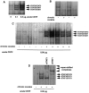
Regulation of STAT DNA binding activity in the VTA by cocaine and CNTF. A, Representative autoradiogram of STAT binding in the VTA 90 min after vehicle (0 μg) or CNTF (0.1 or 0.5 μg) was infused into this brain region of control rats. Four animals were used at each dose with equivalent results.B, Representative autoradiogram of STAT binding in the VTA under basal conditions (i.e., in the absence of intra-VTA infusions) in chronic saline (−)- or chronic cocaine (+)-treated rats. The figure shows an overexposed autoradiogram; there was no consistent effect of cocaine on the intensity of the very low levels of STAT binding apparent under basal conditions. C, Representative autoradiogram of STAT binding in the VTA 90 min after CNTF (0.08 μg) was infused into this brain region of chronic saline (−)- or chronic cocaine (+)-treated rats. Nine animals were used in each treatment group with equivalent results in two separate experiments. The figure illustrates the type of inter-animal variability seen in the magnitude of STAT binding induced by CNTF in chronic cocaine-treated rats. D, Representative autoradiogram of STAT binding analyzed by supershift assays. VTA extracts from a chronic saline (−)- and a chronic cocaine (+)-treated rat from C were used. The identification of the bands as STAT3 homodimers, STAT3:1 heterodimers, or STAT1 homodimers is based on the ability of anti-STAT3 (αSTAT3) and anti-STAT1 (αSTAT1) antibodies to supershift the various bands (see Materials and Methods).
Fig. 4.
Fig. 4.
Regulation of c-Fos and related proteins in the VTA by cocaine and CNTF treatments. A, Representative autoradiograms of c-Fos (58 kDa) and related proteins in the VTA 3 hr after vehicle or CNTF (0.5 μg) was infused into this brain region of control rats. Four animals were used in each treatment group with equivalent results. B, Representative autoradiograms of c-Fos and related proteins in the VTA 3 hr after CNTF (0.1 μg) was infused into this brain region of chronic saline-treated and chronic cocaine-treated rats. Four animals were used in each treatment group with equivalent results. The specificity of the resulting immunoreactivity was demonstrated by its blockade by preabsorption of the antibody with purified M-peptide antigen (Hope et al., 1994).
Fig. 5.
Fig. 5.
Localization of JAK2 within TH- and GFAP-containing cells in the VTA using double-labeling immunohistochemical techniques. A and Bshow representative pictures of JAK2 (red-yellow) colocalized within TH-containing neurons (green). C andD show representative pictures of JAK2 (red-yellow) colocalized within GFAP-containing astrocytes (green). Sections were obtained from chronic cocaine-treated (B, D) and chronic saline-treated (A, C) animals. InC and D, double-labeled cells are indicated by white arrowheads. The results are representative of the analysis of multiple sections of five rats in each treatment group. Note that although the size of JAK2 immunolabeled cells in general appears larger under cocaine-treated conditions, no conclusion about cell size is possible with the methodologies that were used. Scale bar, 25 μm.

References

    1. Angel P, Karin M. The role of Jun, Fos and AP-1 complex in cell-proliferation and transformation. Biochim Biophys Acta. 1991;1072:129–157. - PubMed
    1. Beitner-Johnson D, Nestler EJ. Morphine and cocaine exert common chronic actions on tyrosine hydroxylase in dopaminergic brain reward regions. J Neurochem. 1991;57:344–347. - PubMed
    1. Beitner-Johnson D, Guitart X, Nestler EJ. Glial fibrillary acidic protein and the mesolimbic dopamine system: regulation by chronic morphine and Lewis-Fischer strain differences in the rat ventral tegmental area. J Neurochem. 1993;61:1766–1773. - PubMed
    1. Berhow MT, Russell DS, Terwilliger RZ, Beitner-Johnson D, Self DW, Lindsay RM, Nestler EJ. Influence of neurotrophic factors on morphine- and cocaine-induced biochemical changes in the mesolimbic dopamine system. Neuroscience. 1995;68:969–979. - PubMed
    1. Berhow MT, Hiroi N, Nestler EJ. Regulation of ERK (extracellular signal regulated kinase), part of the neurotrophin signal transduction cascade, in the rat mesolimbic dopamine system by chronic exposure to morphine or cocaine. J Neurosci. 1996;16:4707–4715. - PMC - PubMed

Publication types

MeSH terms