. 1996 Dec 15;16(24):8092-104.
doi: 10.1523/JNEUROSCI.16-24-08092.1996.

Costorage and corelease of modulatory peptide cotransmitters with partially antagonistic actions on the accessory radula closer muscle of Aplysia californica

Affiliations

Costorage and corelease of modulatory peptide cotransmitters with partially antagonistic actions on the accessory radula closer muscle of Aplysia californica

F S Vilim et al. J Neurosci. .

Abstract

Many neurons that contain a classical neurotransmitter also contain modulatory peptides, but it has been difficult to establish unequivocally that these peptides are functional cotransmitters. Here, we provide evidence for functional cotransmission in a neuromuscular system of Aplysia. Using immunocytochemical techniques, we localize members of two peptide families, the small cardioactive peptides (SCPs) and the buccalins (BUCs), to a single subset of dense-core vesicles in the terminals of the cholinergic motorneuron B15. We describe a new preparation and method for the direct detection of released peptides and show that the SCPs and BUCs are released when neuron B15 is intracellularly stimulated. Consistent with their subcellular localization, the SCPs and BUCs are released in a stoichiometric ratio that is constant across conditions that change the absolute amount of peptides released. Peptide release is calcium-dependent but does not require muscle contractions. Thus, the release cannot be attributed to a displacement of peptides that may be present in the extracellular space. In previous studies, we characterized the physiological firing patterns of neuron B15. Here, we simulate these firing patterns and show that peptide release occurs. Additionally, we find that significant quantities of material are released under behaviorally relevant conditions. We find that concentrations of released peptides in the muscle are in the concentration range in which exogenously applied peptides exert characterized modulatory actions on muscle contractions. Together, our findings provide strong support for the hypothesis that peptides contained in neuron B15 are functional cotransmitters.


Figures
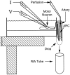
Fig. 1.
Fig. 1.
Diagram of the preparation used to measure peptide release within the ARC. The buccal ganglion is bathed in high-Mg2+ ASW to prevent spontaneous neuronal activity. The motorneuron is impaled with two microelectrodes (one to measure voltage, the other to inject current). These conditions enable precise control over the firing of the motorneuron and ensure that only the motorneuron being fired is responsible for the peptide released within the ARC. The ARC (medium gray) is suspended outside the dish and encased in silicone grease (light gray) and parafilm to prevent dehydration. The nerve connecting the buccal ganglion to the ARC runs through a slit in the side of the dish that is subsequently filled with silicone grease to prevent leakage of solutions from the dish. The ARC is perfused via an artery midway along its length, and the perfusate is collected in drops directly into the tubes that are subsequently used for the measurement of their peptide content. The temperature and length of the ARC were also measured with a temperature probe mounted on an isotonic force transducer (not shown). The tip of the temperature probe was inserted at the bottom of the loop between the base muscle (dark gray) and the artery.
Fig. 2.
Fig. 2.
Cobalt backfill of a buccal ganglion from the ARC muscle. The arrow points to two neurons in the rostral aspect of the ventral motorneuron cluster stained for cobalt. The size and position of the neurons are consistent with the larger one being B15 and the smaller one being B16. Scale bar, 0.5 mm. This agrees with the electrophysiological data indicating that the ARC is innervated by only two motorneurons.
Fig. 3.
Fig. 3.
Buccalin and SCP immunostaining in whole mounts of buccal ganglion and ARC muscle. SCP was stained with a primary antibody generated in the rat and a lissamine rhodamine-conjugated, donkey anti-rat secondary antibody. Buccalin was stained using a primary antibody generated in the rabbit and a fluorescein-conjugated, donkey anti-rabbit secondary antibody. A1 shows buccalin immunostaining of the buccal hemiganglion with the commissure on the right, and A2 shows SCP immunostaining of the same field. The black arrowheads point to a neuron staining for buccalin and SCP, which is of a size and position to be B15. No other cells in the buccal ganglion exhibited costaining for buccalin and SCP. White arrowheads point to a neuron staining for buccalin without staining for SCP, which is of a size and position to be B16. B1 shows buccalin immunostaining of the ARC, andB2 shows SCP immunoreactivity of the same field. Theblack arrowheads point to processes staining for buccalin and SCP, indicating that they belong to B15. White arrowheads point to processes staining for buccalin alone, indicating that they belong to B16. Notice that the B15 and B16 processes are often separated by many micrometers and seem to innervate spatially distinct areas of the ARC. Scale bar (shown inB2), 200 μm.
Fig. 4.
Fig. 4.
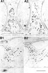
Postembedding immunogold labeling of buccalin and SCP in a B15 neuromuscular junction of the ARC. Cross section of a muscle fiber and B15 terminal in the ARC stained for buccalin (10 nm gold) and SCP (5 nm gold). The presence of buccalin and SCP staining identifies this as a B15 terminal. Note that peptide staining is over the DCVs, not the SCVs, and that many DCVs stain for both buccalin and SCP. The arrow points to an active zone with a number of docked SCVs; note the absence of postsynaptic specializations.f, Myofibrillar bundle in the muscle fiber;g, capping glial process; m, mitochondrion in B15 terminal; c, small clear synaptic vesicles; d, large dense-core vesicles. Scale bar, 0.5 μm.
Fig. 5.
Fig. 5.
Adsorption controls for immunogold labeling of SCP and buccalin in B15 terminals of the ARC. A1 is a cross section stained for SCP (10 nm gold) and buccalin (5 nm gold) showing that many DCVs costain for both. A2 is a serial cross section of the same process as A1 stained in parallel, except that the buccalin primary antibody was pre-adsorbed with synthetic buccalin showing that only the SCP staining remains (10 nm gold). B1 is a longitudinal section stained for SCP (10 nm gold) and buccalin (5 nm gold), also showing that many DCVs costain for both. B2 is a serial longitudinal section of the same process as B1 stained in parallel, except that the SCP primary antibody was adsorbed with synthetic SCP, showing that only the buccalin staining remains (5 nm gold). Scale bar (shown inB2): 0.5 μm.
Fig. 6.
Fig. 6.
Summary of data obtained from immunogold double labeling of B15 terminals in the ARC. Bars represent the percentage of dense-core vesicles that stained for the peptide indicated below the bar. A dense-core vesicle was considered labeled if a gold particle was within 15 nm of its perimeter. The total number of DCVs used to generate the graph is indicated on the y-axis. A1, Rabbit anti-buccalin and rat anti-SCP staining resulted in double labeling of about half of the DCVs. A2, Rabbit anti-SCP and rat anti-SCP staining also resulted in double labeling of about half of the DCVs, suggesting that differences in antibody potency, not differential localization, account for the staining pattern in A1. Also note that >90% of DCVs stain for SCP. B1, Omission or adsorption (with synthetic SCP) of the primary antibody to SCP results in disappearance of the corresponding label. Note that >90% of dense-core vesicles stain for buccalin. B2, Omission or adsorption (with synthetic buccalin) of the primary antibody to buccalin results in the disappearance of the corresponding label. These results indicate that SCP and buccalin are costored in all of the DCVs of B15.
Fig. 7.
Fig. 7.
RIA standard curves for SCP and buccalin. The curves were generated with 125I-labeled peptide, peptide antibody, and serial dilutions of peptide standard. Femtomoles of unlabeled peptide are plotted against the percentage of counts bound to antibody. Zero peptide added was taken as 100% bound and typically accounted for approximately half of the total counts. Each data point is the average ± SE of five separate standard curves generated over a 5 month period. The SCP assay used a 1:5000 dilution of antibody that bound 55.50 ± 0.90% of the counts after a 2 d incubation. The SCP assay had an IC50 of 11.04 ± 0.46 fmol. The buccalin assay used a 1:10,000 dilution of antibody that bound 65.96 ± 0.75% of the counts after a 2 d incubation. The buccalin assay had an IC50 of 18.38 ± 1.29 fmol. The RIAs are quite sensitive and are capable of detecting <1 fmol of peptide.
Fig. 8.
Fig. 8.
Corelease of SCP and buccalin within the ARC in response to stimulation of motorneuron B15. During the period indicated by the bar, the neuron was fired at 12 Hz for 3.5 sec every 7 sec, which is within the physiological range of B15 firing. Samples of ARC perfusate were collected every 2.5 min and analyzed by RIA for their peptide content. Alternate samples were analyzed, respectively, for their SCP or buccalin content. A1, SCP and buccalin release in a single experiment. A2, Same asA1, except that buccalin was scaled by multiplying each buccalin measurement by 2.6, the ratio of total SCP measured divided by the total buccalin measured. With this method, the total SCP and buccalin were taken as identical, enabling a more direct comparison of SCP and buccalin profiles. B1, SCP and buccalin release from four experiments was expressed as a percentage of the total amount of peptide (SCP + buccalin) released in the experiment, and the percentages were averaged for each 5 min period. B2, Same as B1, except that buccalin was scaled so that total percentages were equal for both SCP and buccalin, enabling a more direct comparison of their profiles. Peptide content of the samples increases after stimulation of B15, reaches a peak, and then declines even though the neuron is still being stimulated. BUC, Buccalin.
Fig. 9.
Fig. 9.
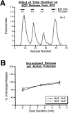
Effect of the total duration of B15 stimulation on peptide release within the ARC. A, Results from a single experiment in which SCP release is measured continuously while periods of stimulation of varying duration are applied; the interburst interval (3.5 sec), the intraburst frequency (12 Hz), and the duration of bursts (3.5 sec) were kept constant. SCP release increases as the period of stimulation lengthens, but the number of action potentials delivered also increases. B, The total released peptide at each of the three durations is corrected to give the release per action potential. This value for each of the three total durations was normalized to the average release for that experiment. The resulting percentage of average release from five separate experiments for each peptide was averaged for each of the three durations. For each peptide, the mean percentage of average release ± SE is plotted against the duration. The results are similar for the two peptides and indicate that there is a slight, but significant, increase in the amount of peptide released per action potential as the duration increases from 5 to 10 min. BUC, Buccalin.
Fig. 10.
Fig. 10.
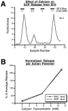
Effect of extracellular calcium concentration on peptide release from B15 in the ARC. A, Results from a single experiment in which SCP release is measured at different calcium concentrations in the perfusate while the intraburst frequency (12 Hz), the burst duration (3.5 sec), the interburst interval (3.5 sec), and the total duration (10 min) are kept constant. SCP release increases with the calcium concentration. B, The total released peptide at each of the calcium concentrations is normalized to the average release for that experiment. The resulting percentage of average release from five separate experiments for SCP, and six separate experiments for buccalin, was averaged for each of the three calcium concentrations. For each peptide, the mean percentage of average release ± SE is plotted against the calcium concentration. The results are similar for the two peptides, indicating that the release of both peptides from B15 is calcium-dependent.BUC, Buccalin.
Fig. 11.
Fig. 11.
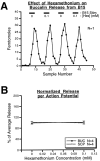
Effect of hexamethonium on peptide release from B15 in the ARC. A, Results from a single experiment in which buccalin release is measured in the presence or absence of 10−4m hexamethonium chloride, which blocks the acetylcholine receptors responsible for generating contraction. The stimulation parameters were all identical. Buccalin release is unaffected by the presence of this ACh antagonist. B, The total released peptide at the two hexamethonium concentrations is normalized using the average release for that experiment. The resulting percentage of average release from four separate experiments for each peptide was averaged for both of the hexamethonium concentrations. For each peptide, the mean percentage of average release ± SE is plotted against the hexamethonium concentration. The results are similar for the two peptides and indicate that peptide release from B15 is unaffected by muscle contraction. BUC, Buccalin.

Similar articles

Cited by

References

    1. Adams ME, O’Shea M. Peptide cotransmitter at a neuromuscular junction. Science. 1983;221:286–289. - PubMed
    1. Andersson PO, Bloom SR, Edwards AV, Jaerhult J. Effects of stimulation of the chorda tympani in bursts on submaxillary response in the cat. J Physiol (Lond) 1982;322:469–483. - PMC - PubMed
    1. Brezina V, Bank B, Cropper EC, Rosen S, Vilim FS, Kupfermann I, Weiss KR. Nine members of the myomodulin family of peptide cotransmitters at the B16-ARC neuromuscular junction of Aplysia. J Neurophysiol. 1995;74:54–72. - PubMed
    1. Church PL, Lloyd PE. Expression of diverse neuropeptide cotransmitters by identified motor neurons in Aplysia. J Neurosci. 1991;11:618–625. - PMC - PubMed
    1. Cohen JL, Weiss KR, Kupfermann I. Motor control of buccal muscles in Aplysia. J Neurophysiol. 1978;41:157–180. - PubMed

Publication types

LinkOut - more resources