. 1996 Dec 15;16(24):8105-14.
doi: 10.1523/JNEUROSCI.16-24-08105.1996.

Release of peptide cotransmitters in Aplysia: regulation and functional implications

Affiliations

Release of peptide cotransmitters in Aplysia: regulation and functional implications

F S Vilim et al. J Neurosci. .

Abstract

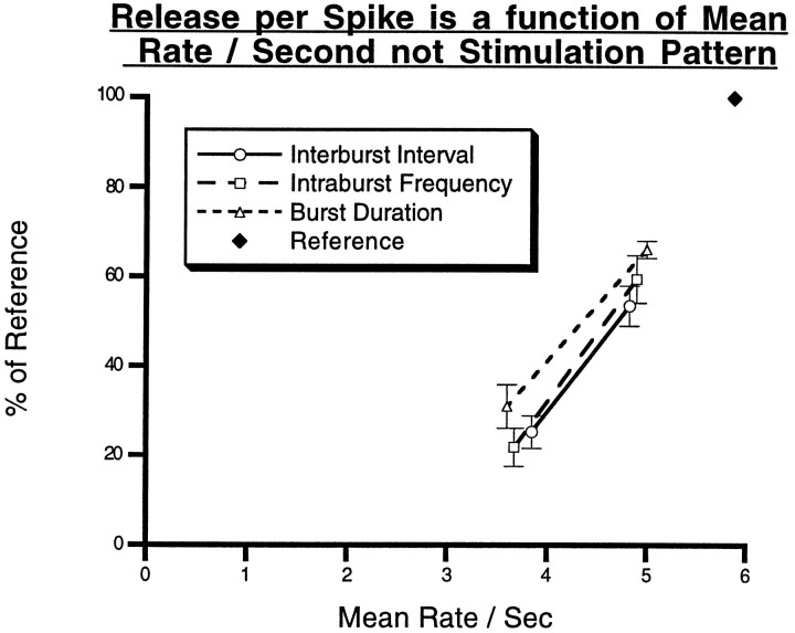
To gain insights into the physiological role of cotransmission, we measured peptide release from cell B15, a motorneuron that utilizes ACh as its primary transmitter but also contains putative peptide cotransmitters, the small cardioactive peptides (SCPs) and the buccalins (BUCs). All stimulation parameters used were in the range in which B15 fires in freely moving animals. We stimulated neuron B15 in bursts and systematically varied the interburst interval, the intraburst frequency, and burst duration. Both peptides were preferentially released when B15 was stimulated at higher intra- or interburst frequencies or with longer burst durations. Across stimulation patterns, the amount of peptide released depended on the mean frequency of stimulation and was independent of the specific pattern of stimulation. The parameters of stimulation that produce a larger release of peptides correspond to those that evoke larger contractions. Large and frequent contractions are likely to fuse or summate, thus disrupting the rhythmic behavior mediated by the muscle innervated by motorneuron B15. Because the combined effect of the SCPs and BUCs is to accelerate the relaxation and shorten the duration of muscle contractions, these peptides reduce the probability of the disruptive fusion or summation of muscle contractions. Because these cotransmitters regulate an aspect of muscle contractions that is not controlled by acetylcholine (ACh), the primary transmitter of B15, we suggest that peptides and ACh form parallel but functionally distinct lines of transmission at the neuromuscular junction. Both types of transmission may be necessary to ensure that behavior remains efficient over a wide range of conditions.


Figures

Fig. 1.
Fig. 1.
The hypothetical role of peptide cotransmitters in the generation of feeding behavior. The solid lines inA and B represent the position of the radula as the animal feeds. Below the dotted line the radula is open, and above thedotted line the radula is closed. In slow feeding, the radula initially closes in response to the contraction of a closer muscle. The closer muscle then relaxes, and the radula is opened by contraction of an opener muscle. The opener then relaxes, and the cycle is repeated. The radula must open and close if functional feeding is to occur. A illustrates what could happen if the rate of feeding is increased while the amplitude and relaxation rate of both opener and closer muscles remains constant. During fast feeding, the contraction of the opener muscle overlaps with that of the closer and, therefore, the opener can no longer provide enough force to open the radula. Radula opening, and therefore functional feeding, can be restored if the closer muscle relaxation rate is increased.B illustrates what happens if the radula closer amplitude is increased with no change in any of the other parameters. Again, the opener muscle is not able to generate enough force to open the radula, and feeding is ineffective. And again, opening and functional feeding can be restored if the closer muscle relaxation rate is increased. This hypothesis predicts that release of peptides should occur when contraction frequency or amplitude is increased.
Fig. 2.
Fig. 2.
Diagram of the systematic changes made in stimulation parameters to test the predictions of the model.A shows the reference stimulation pattern (12 Hz, 3.5 sec on, 3.5 sec off) for motorneuron B15. The reference is identical for all three manipulations. B illustrates the alteration of the IBI without changes in the IF or BD. Cillustrates the alteration of the IF without changes in the IBI or BD.D illustrates alteration of the BD without changes in the IBI or IF. The difference in the total number of action potentials between the reference and the patterns shown inBD is compensated for by expressing peptide release per action potential.
Fig. 3.
Fig. 3.
Effect of IBI on peptide release from B15.A, Results from a single experiment in which SCP release is measured at different IBIs while the IF (12 Hz) and the duration of bursts (3.5 sec) are kept constant. SCP release decreases as the interval between bursts increases. B, The total released peptide at each of the three IBIs is corrected to give the release per action potential. The release per action potential for each of the three IBIs was normalized according to the average release for that experiment. The resulting percentage of average release from five separate experiments for each peptide was averaged for each of the three IBIs. For each of the peptides, the mean percent of average release ± SE is plotted against the IBI. The results are similar for the two peptides and indicate that the amount of peptide released per action potential increases (p < 0.005) as the duration between bursts decreases. The physiological range of IBIs for B15 varies from 3.5 to 10 sec (or more).
Fig. 4.
Fig. 4.
Effect of IF on peptide release from B15.A, Results from a single experiment in which SCP release is measured at three different intraburst frequencies while the duration of bursts (3.5 sec) and the duration between bursts (3.5 sec) are kept constant. SCP release is lower at the lower intraburst frequencies. B, The total released peptide for each of the three frequencies corrected to give the release per action potential. The release per action potential for each of the three frequencies was then normalized according to the average release for that experiment. The resulting percentage of average release from five separate experiments for each peptide was averaged for each of the three frequencies. For each peptide, the mean percent of average release ± SE is plotted against the IF of stimulation. The results are almost identical for the two peptides and indicate that the amount of peptide released per action potential increases (p < 0.005) as the frequency of action potentials increases. The physiological range of firing frequencies for B15 varies between 7.5 and 12 Hz.
Fig. 5.
Fig. 5.
Peptide release is dependent on the BD.A, Results from a single experiment in which SCP release is measured at different BDs while the IF (12 Hz) and the duration between bursts (3.5 sec) are kept constant. SCP release decreases as the BD is reduced. B, The total released peptide for each of the BDs was corrected to give the release per action potential. The release per action potential for each of the three BDs was then normalized according to the average release for that experiment. The resulting percentage of average release from four separate experiments for each peptide was averaged for each of the three BDs. For each peptide, the mean percent of average release ± SE is plotted against the BD. The results are almost identical for the two peptides and indicate that the amount of peptide released per action potential increases (p < 0.005) as the duration of the burst increases. The physiological range of BD varies from 2 to 4 sec for B15.
Fig. 6.
Fig. 6.
Effect of temperature on peptide release from B15.A, Results from a single experiment in which BUC release is measured at three different temperatures while stimulation parameters are kept constant. The BUC release decreases as the temperature is increased. B, The peptide released or the contraction amplitude at each temperature within an experiment was normalized to the average release or average contraction amplitude of that experiment. The resulting percentage of average release from eight separate experiments, four for BUC and four for SCP, was averaged for each peptide at each of the three temperatures and plotted against the temperature. The results are similar for the two peptides: they indicate that the amount of peptide released per action potential increases (p < 0.005) as the temperature decreases. The percent of average contraction amplitude from six separate experiments was averaged and plotted against the temperature. Peptide release and contraction amplitude show a remarkably similar relationship with temperature. The animals are normally housed at 15°C.
Fig. 7.
Fig. 7.
Effect of serotonin and MCC stimulation on peptide release from B15. A, The results from a single experiment in which B15 was stimulated (10 Hz, 3.5 sec on, 3.5 sec off) for four 10 min periods and 5 × 10⁻⁷mserotonin was present in the perfusate in the first and third stimulation period. B, SCP release in a single experiment in which the MCC was stimulated (5 Hz, 3.5 sec on, 3.5 sec off, 10 min duration) along with B15 (10 Hz, 3.5 sec on, 3.5 sec off, 10 min duration) in the second and alone in the fourth stimulation period.
Fig. 8.
Fig. 8.
SCP and serotonin immunostaining in whole mounts of ARC muscle. A shows an optical section of an ARC whole mount stained for SCP with a rat antibody and a fluorescein-conjugated donkey anti-rat secondary antibody.B shows the same optical section stained for serotonin using a rabbit antibody and lissamine rhodamine-conjugated donkey anti-rabbit secondary antibody. Some, but not all, SCP-staining varicosities have serotonin staining associated with them, indicating that MCC processes fasciculate with B15 processes and that there may be an opportunity for presynaptic facilitation of transmitter release in some of the B15 terminals. Scale bar, 100 μm.
Fig. 9.
Fig. 9.
Effect of BUC A on SCP release from B15.A, SCP release in a single experiment in which 5 × 10⁻⁶m BUC A was present in the perfusate in the third stimulation period. The stimulation parameters were kept constant in each of the four 10 min stimulation periods (black bars). B, The combined results from five experiments in which release is expressed as a percentage of the average release from the periods before, during, and after the application of the BUC A. The bars represent mean ± SE for each period. BUC produces a significant (p < 0.01) decrease in SCP release that reverses with washout.
Fig. 10.
Fig. 10.
The data from the stimulation parameter experiments (IBI, IF, and BD) were re-analyzed by comparing the release in each experiment to the common reference conditions (12 Hz, 3.5 sec on, 3.5 sec off). Because all of the experiments had the common reference conditions, the results of different experiments could be directly compared. The data were expressed as percent of the release in the reference condition, averaged for like stimulation conditions, and plotted as a function of the average frequency (total action potentials delivered/total seconds) for that condition. In this way, the results from all three stimulation paradigms (IBI, IF, and BD) could be plotted on common axes and compared. The significant overlap in the graphs of the three stimulation paradigms indicates that peptide release is more dependent on the average frequency (mean rate/sec) than on the way in which this average frequency was achieved, at least within the range that is observed in vivo. The black diamond indicates the reference stimulation condition that is shared by all three stimulation paradigms and, consequently, all three lines converge at this point. Extrapolation of these lines to thex-axis indicates that they cross at ∼3 Hz, suggesting that this is the threshold average frequency for the peptide release from motorneuron B15.

Similar articles

Cited by

References

    1. Adams ME, O’Shea M. Peptide cotransmitter at a neuromuscular junction. Science. 1983;221:286–289. - PubMed
    1. Andersson PO, Bloom SR, Edwards AV, Jaerhult J. Effects of stimulation of the chorda tympani in bursts on submaxillary response in the cat. J Physiol (Lond) 1982;322:469–483. - PMC - PubMed
    1. Brezina V, Evans CG, Weiss KR. Enhancement of Ca current in the accessory radula closer muscle of Aplysia californica by neuromodulators that potentiate its contractions. J Neurosci. 1994;14:4393–4411. - PMC - PubMed
    1. Cohen JL, Weiss KR, Kupfermann I. Motor control of buccal muscles in Aplysia. J Neurophysiol. 1978;41:157–180. - PubMed
    1. Cropper EC, Lloyd PE, Reed W, Tenenbaum R, Kupfermann I, Weiss KR. Multiple neuropeptides in cholinergic motor neurons of Aplysia: evidence for modulation intrinsic to the motor circuit. Proc Natl Acad Sci USA. 1987;84:3486–3490. - PMC - PubMed

Publication types

LinkOut - more resources