. 1996 Dec 15;16(24):8132-9.
doi: 10.1523/JNEUROSCI.16-24-08132.1996.

GDNF selectively protects dopamine neurons over serotonin neurons against the neurotoxic effects of methamphetamine

Affiliations

GDNF selectively protects dopamine neurons over serotonin neurons against the neurotoxic effects of methamphetamine

W A Cass. J Neurosci. .

Abstract

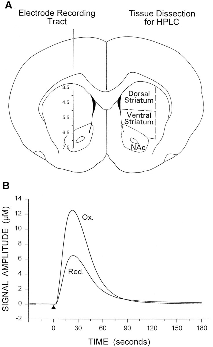
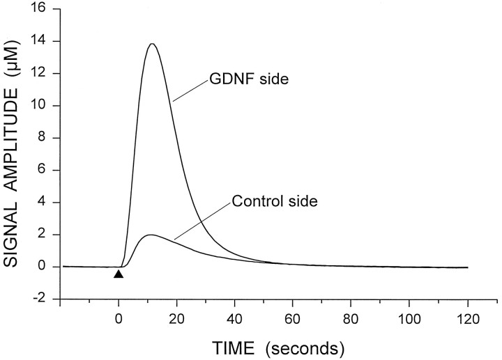
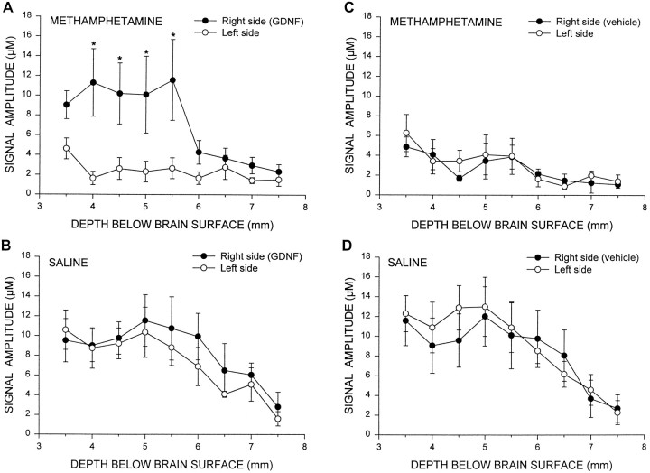
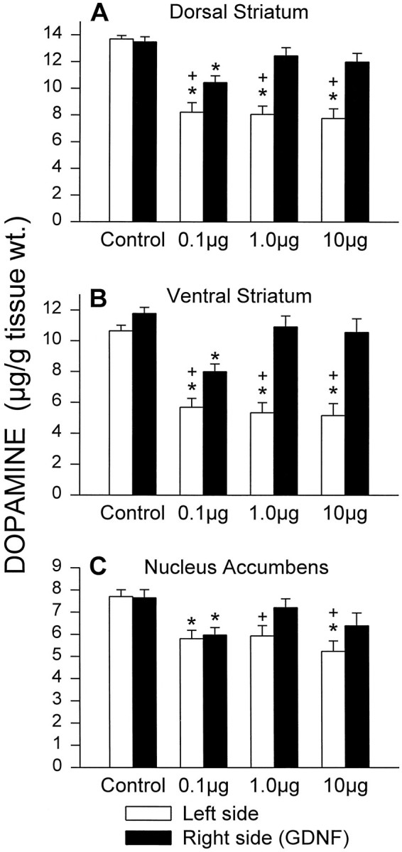
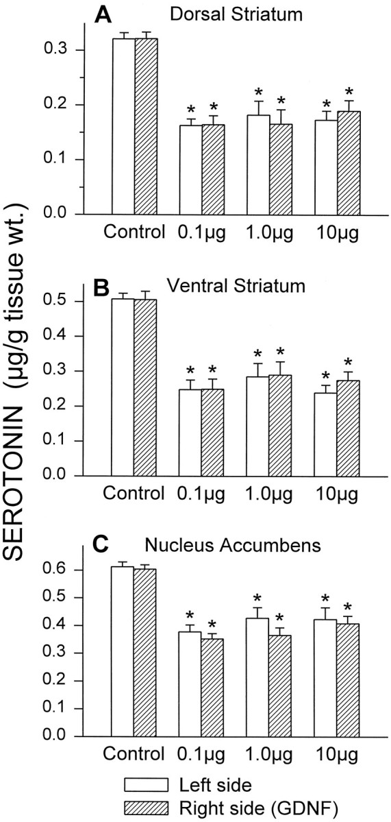
Repeated methamphetamine (METH) administration to animals can result in long-lasting decreases in striatal dopamine (DA) and serotonin (5-HT) levels. Glial cell line-derived neurotrophic factor (GDNF) has pronounced effects on dopaminergic systems in vivo, including partial neuroprotective effects against 6-hydroxydopamine and 1-methyl-4-phenyl-1,2,3,6-tetrahydropyridine -induced lesions. The present study examined the ability of GDNF to prevent METH-induced reductions in potassium-evoked overflow of DA, and DA and 5-HT content, in striatum. GDNF (10 microg) or vehicle was injected into the right striatum of anesthetized rats. Twenty-four hours later, the rats were injected four times at 2 hr intervals with METH (5 mg/kg, s.c.) or saline. One week later, in vivo electrochemistry was used to monitor the overflow of DA evoked by local potassium application. Evoked overflow of DA was dramatically decreased in the striatum of METH-treated animals. GDNF prevented the reduction in evoked overflow of DA in the right striatum of the METH-treated animals. After each experiment, the animals were killed, and striatal DA and 5-HT levels determined by HPLC. The METH treatment produced significant decreases in both neurotransmitters. GDNF administration prevented the reduction in striatal DA levels on the treated side of the brain, whereas levels on the contralateral side were still decreased. In dose-response studies, 1 microg of GDNF was as protective as 10 microg, whereas 0.1 microg was only partially protective. In contrast, 5-HT levels were only minimally protected by previous administration of GDNF. These results suggest that GDNF can selectively protect DA neurons, compared with 5-HT neurons, against the neurotoxic effects of METH.


Figures

Fig. 1.
Fig. 1.
A, Illustration of the position of the recording electrode track for the in vivoelectrochemistry experiments and the dissection of the striatum and nucleus accumbens (NAc) for HPLC analysis of monoamines. This coronal section is ∼1.2 mm rostral to bregma (Paxinos and Watson, 1986). B, Representative signal showing the potassium-evoked overflow of DA in the striatum of a control rat. Potassium (300 nl, 70 mm K+) was applied at thearrowhead. The oxidation (Ox.) and reduction (Red.) current responses (reduction/oxidation current ratio = 0.52) indicate that the predominate electroactive species detected is DA.
Fig. 2.
Fig. 2.
Representative signals for the potassium-evoked overflow of DA from the right side (GDNF side) and left side (Control side) of the dorsal striatum of a GDNF- and METH-treated animal. Potassium solution (250 nl) was applied at thearrowhead in each case. For clarity, only the oxidation signals are shown.
Fig. 3.
Fig. 3.
Summary of potassium-evoked DA signal amplitude throughout the striatum and nucleus accumbens of GDNF-treated animals administered METH (A) or saline (B) and vehicle-treated animals administered METH (C) or saline (D). GDNF or vehicle was injected into the right striatum 24 hr before METH or saline treatment. In vivoelectrochemical recordings were made 1 week after METH or saline administration. The data shown are mean ± SEM values for six animals per group for the GDNF groups (A,B) and five animals per group for the saline groups (C, D). The data were analyzed using two-factor ANOVA with side of brain and depth of recording as within factors. F scores for the GDNF and METH group (A): side F = 12.33,p = 0.025; depth F = 3.32,p = 0.007; interaction F = 3.32, p = 0.007. F scores for the GDNF and saline group (B): side F = 0.74, p = 0.439; depth F = 11.46, p < 0.001; interactionF = 1.47, p = 0.206.F scores for the vehicle and METH group (C): side F = 0.45,p = 0.540; depth F = 4.20,p = 0.002; interaction F = 0.74, p = 0.658. F scores for the vehicle and saline group (D): side F= 0.36, p = 0.579; depth F = 5.05, p < 0.001; interaction F= 0.82, p = 0.591. *p < 0.05 versus left side at same depth (Newman–Keuls post hoccomparisons).
Fig. 4.
Fig. 4.
DA levels in the striatum and nucleus accumbens of GDNF-treated animals administered METH 24 hr after the GDNF. Doses of GDNF (0.1, 1.0, and 10 μg) are shown on the horizontal axis. Control animals were given intrastriatal injections of vehicle and administered saline 24 hr later. Tissue was taken 1 week after METH or saline treatment and divided into dorsal striatum (A), ventral striatum (B), and nucleus accumbens (C), as indicated in Figure1A. The data shown are mean ± SEM values for 9 or 10 animals per group. The data were analyzed using two-factor ANOVA with side of brain as a within factor. F scores for dorsal striatum (A): dose F = 12.0, p < 0.001; side F = 109.2, p < 0.001; interactionF = 17.6, p < 0.01.F scores for ventral striatum (B): doseF = 8.67, p < 0.001; sideF = 202.8, p < 0.001; interaction F = 13.6, p < 0.001. F scores for nucleus accumbens (C): dose F = 5.10,p = 0.005; side F = 13.7,p < 0.001; interaction F = 3.86, p = 0.02. *p < 0.05 versus same side of control group; +p< 0.05 versus right side of brain at same dose (Newman–Keulspost hoc comparisons).
Fig. 5.
Fig. 5.
5-HT levels in the striatum and nucleus accumbens of GDNF-treated animals administered METH 24 hr after the GDNF. Doses of GDNF (0.1, 1.0, and 10 μg) are shown on the horizontal axis. Control animals were given intrastriatal injections of vehicle and administered saline 24 hr later. Tissue was taken 1 week after METH or saline treatment and divided into dorsal striatum (A), ventral striatum (B), and nucleus accumbens (C), as indicated in Figure1A. The data shown are mean ± SEM values for 9 or 10 animals per group. The data were analyzed using two-factor ANOVA with side of brain as a within factor. F scores for dorsal striatum (A): dose F = 16.5, p < 0.001; side F = 0.08, p = 0.78; interaction F = 2.18, p = 0.11. F scores for ventral striatum (B): dose F = 16.7,p < 0.001; side F = 0.78,p = 0.38; interaction F = 0.94,p = 0.43. F scores for nucleus accumbens (C): dose F = 20.4,p < 0.001; side F = 3.14,p = 0.09; interaction F = 0.59,p = 0.63. *p < 0.05 versus same side of control group (Newman–Keuls post hoccomparisons).

References

    1. Axt KJ, Mamounas LA, Molliver ME. Structural features of amphetamine neurotoxicity in the brain. In: Cho AK, Segal DS, editors. Amphetamine and its analogs: psychopharmacology, toxicology, and abuse. Academic; San Diego: 1994. pp. 315–367.
    1. Beck KD, Valverde J, Alexi T, Poulsen K, Moffat B, Vandlen RA, Rosenthal A, Hefti F. Mesencephalic dopaminergic neurons protected by GDNF from axotomy-induced degeneration in the adult brain. Nature. 1995;373:339–341. - PubMed
    1. Beck KD, Irwin I, Valverde J, Brennan TJ, Langston JW, Hefti F. GDNF induces a dystonia-like state in neonatal rats and stimulates dopamine and serotonin synthesis. Neuron. 1996;16:665–673. - PubMed
    1. Bittner SE, Wagner GC, Aigner TG, Seiden LS. Effects of high-dose treatment of methamphetamine on caudate dopamine and anorexia in rats. Pharmacol Biochem Behav. 1981;14:481–486. - PubMed
    1. Bowenkamp KE, Hoffman AF, Gerhardt GA, Henry MA, Biddle P, Hoffer BJ, Granholm A-CE. Glial cell line-derived neurotrophic factor supports survival of injured midbrain dopaminergic neurons. J Comp Neurol. 1995;355:479–489. - PubMed

Publication types