Comparative Study
. 1997 Jan 7;94(1):48-52.
doi: 10.1073/pnas.94.1.48.

Spare the rod, spoil the regulation: necessity for a myosin rod

Affiliations
Comparative Study

Spare the rod, spoil the regulation: necessity for a myosin rod

K M Trybus et al. Proc Natl Acad Sci U S A. .

Abstract

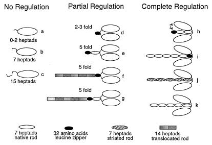
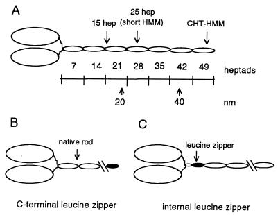
Regulation of a variety of cellular contractile events requires that vertebrate smooth and non-muscle myosin II can achieve an "off" state. To examine the role of the myosin rod in this process, we determined the minimal size at which a myosin molecule is capable of regulation via light chain phosphorylation. Expressed smooth muscle myosin subfragments with as many as 100 amino acids of the coiled-coil rod sequence did not dimerize and were active independently of phosphorylation. To test whether dimerization per se restores regulation of ATPase activity, mutants were expressed with varying lengths of rod sequence, followed by C-terminal leucine zippers to stabilize the coiled-coil. Dimerization restored partial regulation, but the presence of a length of rod approximately equal to the myosin head was necessary to achieve a completely off state. Partially regulated short dimers could be converted into fully regulated molecules by addition of native rod sequence after the zipper. These results suggest that the myosin rod mediates specific interactions with the head that are required to obtain the completely inactive state of vertebrate smooth and non-muscle myosins. If these interactions are prohibited under cellular conditions, unphosphorylated crossbridges can slowly cycle.


Figures

Figure 4
Figure 4
Schematic diagram grouping the constructs that show no regulation, partial regulation (<10-fold), or complete regulation (>10-fold) by light chain phosphorylation as determined by steady-state actin-activated ATPases. All monomeric constructs (a–c), regardless of length, show no regulation with phosphorylation. Constructs that show partial regulation include C-terminal zippers after 0, 2, or 7 heptads of native sequence (d and e), and constructs in which a piece of rod is translocated from its normal position relative to the invariant proline (f and g). Among the constructs showing full regulation are 15-heptad/zipper (h), a 37-heptad construct with the leucine zipper replacing amino acids 867–898 (i), a smooth head/striated rod chimera (j), and a 37-heptad wild-type construct (k). Dimers that are fully regulated are shown with their heads bent down toward the rod to indicate that specific sequences in the rod mediate head–rod or head–head interactions. The head–rod interaction is likely to be most stable in 10S myosin, which has the lowest rate of product release. The changes in head disposition that occur in HMM may be more subtle than implied by this diagram, which was drawn to emphasize a head–rod interaction.
Figure 1
Figure 1
(A) Schematic diagram of double-headed heavy meromyosin. An invariant proline (amino acid 849 in the gizzard heavy chain) marks the end of the head and the beginning of the heptad repeat in the rod. Each oval-shaped segment of the rod represents 7 heptad repeats. One heptad repeat has a repeated pattern of seven amino acids (abcdefg), where the a and d positions have a preponderance of hydrophobic residues (11). The rise per residue is 0.15 nm. CHT-HMM, HMM prepared by chymotryptic digestion of myosin. (B) Illustration of the C-terminal leucine zipper constructs. The C-terminal leucine zipper constructs are named for the number of heptads of native sequence before the leucine zipper sequence, e.g., 2-heptad/zipper has 2 heptads of rod sequence followed by the 32-aa leucine zipper sequence. (C) Illustration of the internal leucine zipper constructs, in which native rod sequence follows the leucine zipper sequence.
Figure 2
Figure 2
(A) Native gel showing the dimerization state of the expressed constructs. Lane 1, dimeric skeletal rod chimera (construct j); lane 2, dimeric 25-heptad short HMM with a C-terminal leucine zipper (similar to construct h); lane 3, 25-heptad short HMM without the zipper, showing an equilibrium between dimeric and monomeric species; lane 4, dimeric 2-heptad construct with a C-terminal leucine zipper (construct d); lane 5, monomeric 15-heptad construct (construct c). (B) Metal-shadowed images of the dimeric 2-heptad construct with a C-terminal leucine zipper (Left; construct d) show pairs of heads (arrowheads), in contrast to the single heads of monomeric S1 (Right).
Figure 3
Figure 3
Actin-activated ATPase activity of monomers and C-terminal leucine zipper constructs. Phosphorylated species, open symbols; unphosphorylated species, filled symbols. (A) Monomeric 15-heptad S1 (construct c). (B) C-terminal 2-heptad/zipper (construct d). (C) C-terminal 7-heptad/zipper (construct e). (D) C-terminal 15-heptad/zipper (circles; construct h) and 25-heptad short HMM/zipper (squares).

Similar articles

Cited by

References

    1. Harrington W F, Rodgers M E, Davis J S. In: Molecular Mechanisms in Muscular Contraction. Squire J M, editor. New York: Macmillan; 1990. pp. 241–263.
    1. Itakura S, Yamakawa H, Toyoshima Y Y, Ishijima A, Kojima T, Harada Y, Yanagida T, Wakabayashi T, Sutoh S. Biochem Biophys Res Commun. 1993;196:1504–1510. - PubMed
    1. Trybus K M. J Muscle Res Cell Motil. 1994;15:587–594. - PubMed
    1. Cremo C R, Sellers J R, Facemyer K C. J Biol Chem. 1995;270:2171–2175. - PubMed
    1. O’Shea E K, Klemm J D, Kim P S, Alber T. Science. 1991;254:539–544. - PubMed

Publication types

MeSH terms

LinkOut - more resources