

The B12-dependent ribonucleotide reductase from the archaebacterium Thermoplasma acidophila: an evolutionary solution to the ribonucleotide reductase conundrum
- PMID: 8990160
- PMCID: PMC19235
- DOI: 10.1073/pnas.94.1.53
The B12-dependent ribonucleotide reductase from the archaebacterium Thermoplasma acidophila: an evolutionary solution to the ribonucleotide reductase conundrum
Abstract
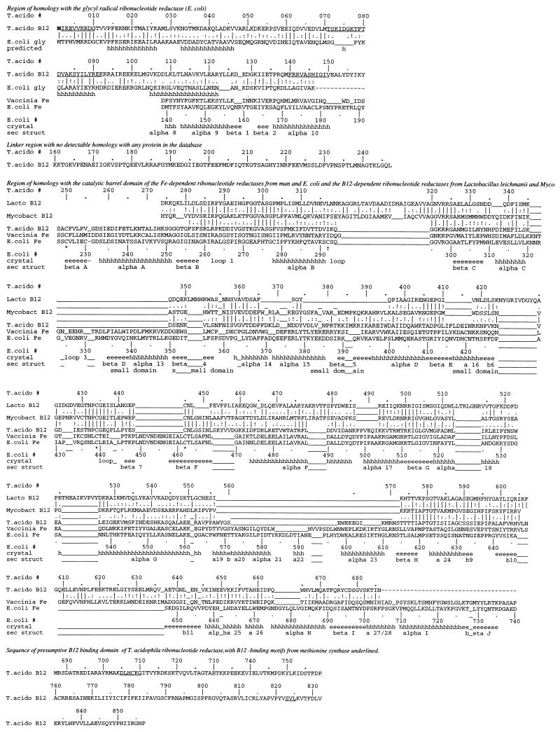
A coenzyme B12-dependent ribonucleotide reductase was purified from the archaebacterium Thermoplasma acidophila and partially sequenced. Using probes derived from the sequence, the corresponding gene was cloned, completely sequenced, and expressed in Escherichia coli. The deduced amino acid sequence shows that the catalytic domain of the B12-dependent enzyme from T. acidophila, some 400 amino acids, is related by common ancestry to the diferric tyrosine radical iron(III)-dependent ribonucleotide reductase from E. coli, yeast, mammalian viruses, and man. The critical cysteine residues in the catalytic domain that participate in the thiyl radical-dependent reaction have been conserved even though the cofactor that generates the radical is not. Evolutionary bridges created by the T. acidophila sequence and that of a B12-dependent reductase from Mycobacterium tuberculosis establish homology between the Fe-dependent enzymes and the catalytic domain of the Lactobacillus leichmannii B12-dependent enzyme as well. These bridges are confirmed by a predicted secondary structure for the Lactobacillus enzyme. Sequence similarities show that the N-terminal domain of the T. acidophila ribonucleotide reductase is also homologous to the anaerobic ribonucleotide reductase from E. coli, which uses neither B12 nor Fe cofactors. A predicted secondary structure of the N-terminal domain suggests that it is predominantly helical, as is the domain in the aerobic E. coli enzyme depending on Fe, extending the homologous family of proteins to include anaerobic ribonucleotide reductases, B12 ribonucleotide reductases, and Fe-dependent aerobic ribonucleotide reductases. A model for the evolution of the ribonucleotide reductase family is presented; in this model, the thiyl radical-based reaction mechanism is conserved, but the cofactor is chosen to best adapt the host organism to its environment. This analysis illustrates how secondary structure predictions can assist evolutionary analyses, each important in "post-genomic" biochemistry.
Figures
References
Publication types
MeSH terms
Substances
LinkOut - more resources
Full Text Sources
