Comparative Study
. 1997 Jan 7;94(1):65-72.
doi: 10.1073/pnas.94.1.65.

Folding of the N-terminal, ligand-binding region of integrin alpha-subunits into a beta-propeller domain

Affiliations
Comparative Study

Folding of the N-terminal, ligand-binding region of integrin alpha-subunits into a beta-propeller domain

T A Springer. Proc Natl Acad Sci U S A. .

Abstract

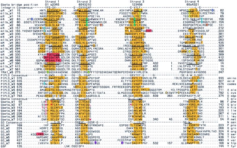
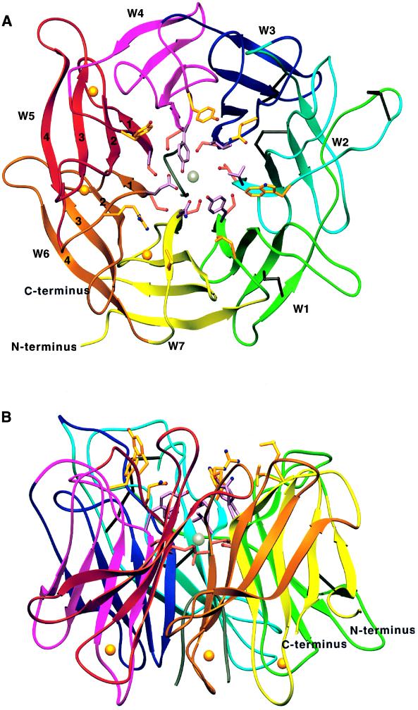
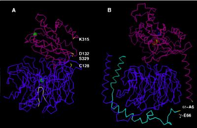
The N-terminal approximately 440 aa of integrin alpha subunits contain seven sequence repeats. These are predicted here to fold into a beta-propeller domain. A homologous domain from the enzyme phosphatidylinositol phospholipase D is predicted to have the same fold. The domains contain seven four-stranded beta-sheets arranged in a torus around a pseudosymmetry axis. The trimeric G-protein beta subunit (G beta) appears to be the most closely related beta-propeller. Integrin ligands and a putative Mg2+ ion are predicted to bind to the upper face of the beta-propeller. This face binds substrates in beta-propeller enzymes and is used by the G protein beta subunit to bind the G protein alpha subunit. The integrin alpha subunit I domain, which is structurally homologous to the G protein alpha subunit, is tethered to the top of the beta-propeller domain by a hinge that may allow movement of the domains relative to one another. The Ca2+-binding motifs in integrin alpha subunits are on the lower face of the beta-propeller.


Figures

Figure 2
Figure 2
Alignments, secondary structure, and disulfide bonds. Residues in human integrin α-subunits and bovine phosphatidylinositol phospholipase D (PIPLD) predicted by PHD to be β-strand and α-helix are highlighted in gold and magenta, respectively. The known secondary structure elements of G beta and GO β-propeller domains are highlighted with the same colors. Residues present in β-strands in the α4 model as determined by dssp (26) are underlined. Cysteines known in αIIb (29) or predicted in α4 and αM to be disulfide bonded together are coded with the same color. Equivalent ladder positions in each strand are numbered at the top for G beta; G beta and GO are structurally aligned. The position of framework residues is marked with a line below the consensus sequence for G beta and GO. Residues in G beta that contact the switch II region of the G α-subunit (37, 44) are shown in red. The alignment unit is the sheet (W) rather than the sequence repeat; note that the N-terminal segment is in strand 4, W7. For structural alignments, the seven β-sheets (Ws) of GO (38) and G beta (24, 37, 44) were cut out and structurally superimposed on one another, and the intact β-propellers were also superimposed using the program 3dmalign (45). Sequence alignments were prepared with pileup in gcg (22) and manually adjusted with megalign (DNAstar, Madison, WI). All gaps except those between strands were removed. Consensus sequences were calculated with pretty in gcg using a plurality of 3/7 of the total number of W.
Figure 1
Figure 1
Folding topology of the integrin β-propeller. The Ws are upright. β-strands are arrows, known disulfides in αIIb (29) are horizontal lines, and boundaries between FG-GAP repeats are marked with vertical dashes.
Figure 3
Figure 3
Ribbon diagrams (65) of the model for the integrin α4-subunit β-propeller domain. Views are from the top (A) and side (B). Each W is shown in a different color. A hypothetical polypeptide finger in the central cavity is gray. Cysteines in disulfides are black. Side chains in β-strand 1 in positions 0, b, and 2 are shown in gold, lavender, and rose, respectively; their oxygens and nitrogens are red and blue, respectively. Ca2+ ions and a hypothetical Mg2+ ion are gold and silver spheres, respectively.
Figure 4
Figure 4
Superimposition of the αM integrin subunit I domain and β-propeller domain on a trimeric G protein. (A) The αM I domain (magenta; ref. 17), β-propeller domain (purple), and polypeptide finger (white) are shown with Cα traces. Mg2+ and Ca2+ ions are green and grey spheres, respectively. Residues at the ends of the domains that may be connected by linker segments are shown in yellow. According to the superimposition, the α carbons of C128 and D132 are 7.4 Å apart, and K315 and S329 are 11 Å apart. (B) The trimeric G protein α-subunit (magenta) and bound GDP (space-filling), β-subunit (purple), and γ-subunit (cyan; ref. 44) are shown in the same orientation. The residues in yellow are separated by 5–7 residues from lipid-modified termini that bind to the inner face of the plasma membrane (37, 44). These interactions with the plasma membrane increase association between the α-subunit and the βγ-subunit dimer and to some extent may act like the covalent linkage between the I domain and β-propeller domain in integrin α-subunits. The αM β-propeller domain was modeled on α4, and superimposed on G beta with the alignment in Fig. 2 and a 2/7 rotation on the pseudosymmetry axis—i.e. with αM W1 on G beta W6. A total of 49 residues in αM I domain elements βA, α1, βB, βD, βE, βF, and α7 (17) was superimposed on Gα β1, α1, β3, β4, β5, β6, and α5 (44), respectively, with an RMS of 1.56 Å using look. The figure was made with look.

References

    1. Springer T A. Nature (London) 1990;346:425–433. - PubMed
    1. Hemler M E. Annu Rev Immunol. 1990;8:365–400. - PubMed
    1. Hynes R O. Cell. 1992;69:11–25. - PubMed
    1. Smyth S S, Joneckis C C, Parise L V. Blood. 1993;81:2827–2843. - PubMed
    1. Dustin M L, Springer T A. Nature (London) 1989;341:619–624. - PubMed

Publication types

LinkOut - more resources