. 1997 Jan 7;94(1):79-84.
doi: 10.1073/pnas.94.1.79.

Accessibility of nuclear DNA to triplex-forming oligonucleotides: the integrated HIV-1 provirus as a target

Affiliations

Accessibility of nuclear DNA to triplex-forming oligonucleotides: the integrated HIV-1 provirus as a target

C Giovannangeli et al. Proc Natl Acad Sci U S A. .

Abstract

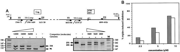
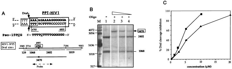
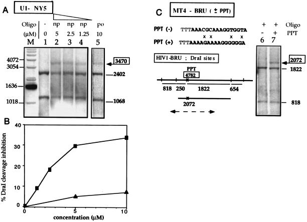
The control of gene transcription by antigene oligonucleotides rests upon the specific recognition of double-helical DNA by triplex-forming oligonucleotides. The development of the antigene strategy requires access to the targeted DNA sequence within the chromatin structure of the cell nucleus. In this sudy we have used HIV-1 chronically infected cells containing the HIV provirus as endogenous genes to demonstrate that the integrated HIV-1 proviral genome is accessible to triplex-forming oligonucleotides within cell nuclei. An oligonucleotide-psoralen conjugate targeted to the polypurine tract (PPT) of the HIV-1 proviral sequence was used as a tool to convert the noncovalent triple-helical complex into a covalent lesion on genomic DNA after UV irradiation of cells. Triplex-derived adducts were analyzed using two different methods. The photo-induced psoralen cross-link prevented cleavage of the target sequence by DraI restriction endonuclease, and the sequence-specific inhibition of cleavage was revealed and quantitated by Southern blot analysis. A quantitative analysis of cross-linking efficiency was also carried out by a competitive PCR-based assay. These two approaches allowed us to demonstrate that a triplex-forming oligonucleotide can recognize and bind specifically to a 15-bp sequence within the chromatin structure of cell nuclei.


Figures

Figure 3
Figure 3
Triplex-induced cross-link detection on genomic DNA by a competitive PCR-based assay. (A Upper) Schematic representation of the genomic HIV region, where the two primer pairs (Gag–SX, 5′-ATAATCCACCTATCCCAGTAGGAGAAAT-3′; and Gag–DX, 5′-TTTGGTCCTTGTCTTATGTCCAGAATGC-3′; PPT–SX, 5′-CCCTACAATCCCCAAAGTCAAGGAGTAA-3′; and PPT-DX, 5′-GATCTCTGCTGTCCCTGTAATAAACCCG-3′) are located (converging arrows). For each primer pair, a competitor DNA fragment for quantitative PCR was constructed using DX primer and the internal SX–Comp primer [Gag–SX–Comp, 5′(Gag–SX)-CCTGGGATTAAATAAAATAG-3′; PPT–SX–Comp, 5′(PPT–SX)-CAGGTAAGAGATCAGGCTGA-3′, broken arrows] and quantitated. Before PCR amplification, genomic DNA was digested by EcoRI and AvaI which cut outside of the amplified regions as indicated. Nucleotide numbers refer to positions in HIV-1–NY5 (GenBank). (A Lower) Competitive PCR experiment for quantitation of the amount of cross-linked DNA molecules. Genomic DNA irradiated with 10 μM of Pso-15TCG(po) (see Fig. 1B, lane 2, which corresponds to the same sample analyzed by DraI protection assay) was quantitated by coamplification of a fixed amount of sample with increasing concentrations of competitor, as indicated near the gels. For each gel the genomic and competitor products are indicated. The results of quantitation obtained for these samples (20000 Gag molecules and 7400 PPT molecules) indicate that 63% of the DNA molecules are cross-linked. Lane M, DNA marker (VIII, Boehringer Mannheim). (B) Comparison of quantitations of the same sample after Pso-15TCG(po) treatment obtained by DraI protection assay (grey bars, corresponding to the samples of the experiment reported on Fig. 1) and by competitive PCR-based assay (white bars).
Figure 1
Figure 1
DraI protection assay on naked genomic DNA. (A Upper) The 16-bp oligopurine·oligopyrimidine sequence we have chosen as a target for triple helix formation is indicated in boldface type. It is present in the HIV-1 genome and is called PPT. The PPT triplex site overlaps the DraI recognition sequence (thin line); the two arrows indicate the sites of DraI cleavage. The sequence of the psoralen-modified third strand is indicated. Underlined cytosine (C) indicates a 5 methyl cytosine. Pso- indicates a psoralen derivative attached to the 5′ phosphate (see Materials and Methods). (A Lower) DraI sites around the PPT triplex site (which is shown by a cross and boxed) present in the pol gene (HIV-NY5 sequence). sequence). The lengths of the fragments obtained after DraI cleavage are indicated. Location of the DNA probe (1902 bp long) used for Southern blot analysis is shown (broken arrow); the probe hybridizes with the 1068- and 2402-bp fragments over 897 and 1005 nt, respectively. (B) Naked genomic DNA (0.8 μg/μl) of U1 cells was irradiated (in the triplex-binding buffer) in the absence (lane 1) and in the presence of different concentrations of Pso-15TCG(po) (lane 2, 10 μM; lane 3, 5 μM; lane 4, 2.5 μM); the genomic DNA was then analyzed by DraI protection assay. Lane M, DNA marker (GIBCO/BRL). Fragments lengths (in base pairs) are indicated near the gel. (C) Quantitation of DraI cleavage inhibition at the PPT triplex site on the naked genomic DNA (as described in Fig. 2B) as a function of oligonucleotide concentration (▪, np; ▴, po). The ratio of the number of counts at the 3470-bp fragment to the sum of the 3470-, 2402-, and 1068-bp fragments are shown.
Figure 2
Figure 2
Triplex formation in nuclei of permeabilized cells. Chronically infected cells were permeabilized with digitonin, incubated in the triplex-forming buffer in the absence or in the presence of Pso-15TCG, and irradiated. After cell lysis, followed by proteinase K and RNase A treatments, the genomic DNA was prepared and analyzed by DraI protection assay. Chronically infected cells were used (U1–NY5, MT4–BRU). They were obtained by infection with different HIV-1 strains (called NY5 and BRU) and differed from each other by the HIV-1 sequence. However the PPT triplex site sequence was the same in all strains except in the MT4–BRU(−) cell line where the PPT sequence was mutated at four positions (see C). These sequence differences led to slightly different DraI cleavage patterns, as indicated under the gels, but even though the fragment lengths were modified from one cell line to the other, specific triplex formation was always correlated with the appearance of a longer fragment of well-defined size; probe location is shown by a broken line for MT4-BRU (see Fig. 1 for U1-NY5). (A) U1 cells (HIV-1–NY5 sequence); the po or np oligomers and the concentration of the Pso-15TCG are shown by the gels. (B) Quantitation of DraI cleavage inhibition at the PPT triplex site in nuclei of permeabilized U1 cells versus oligonucleotide concentration (np, ▪; po, ▴). (C) MT4 cells [HIV-1–BRU(+), lane 7; or HIV-1-BRU(−), lane 6] treated with Pso-15TCG(po). The sequence of the mutated PPT [PPT(−)] is indicated and mismatches with the wild PPT [PPT(+)] are indicated by crosses (×).

References

    1. Thuong N T, Hélène C. Angew Chem Int Ed Engl. 1993;32:666–690.
    1. Maher L J., III Cancer Invest. 1996;14:66–82. - PubMed
    1. Grigoriev M, Praseuth D, Guieysse A L, Robin P, Thuong N T, Hélène C, Harel-Bellan A. Proc Natl Acad Sci USA. 1993;90:3501–3505. - PMC - PubMed
    1. Roy C. Nucleic Acids Res. 1993;21:2845–2852. - PMC - PubMed
    1. Degols G, Clarenc J P, Lebleu B, Léonetti J P. J Biol Chem. 1994;269:16933–16937. - PubMed

Publication types

MeSH terms

LinkOut - more resources