. 1997 Jan 7;94(1):85-90.
doi: 10.1073/pnas.94.1.85.

Drosophila TAF(II)230 and the transcriptional activator VP16 bind competitively to the TATA box-binding domain of the TATA box-binding protein

Affiliations

Drosophila TAF(II)230 and the transcriptional activator VP16 bind competitively to the TATA box-binding domain of the TATA box-binding protein

J Nishikawa et al. Proc Natl Acad Sci U S A. .

Abstract

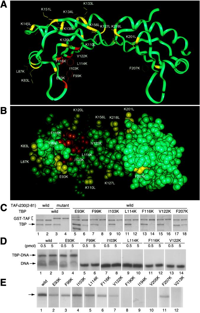
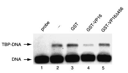
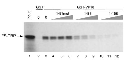
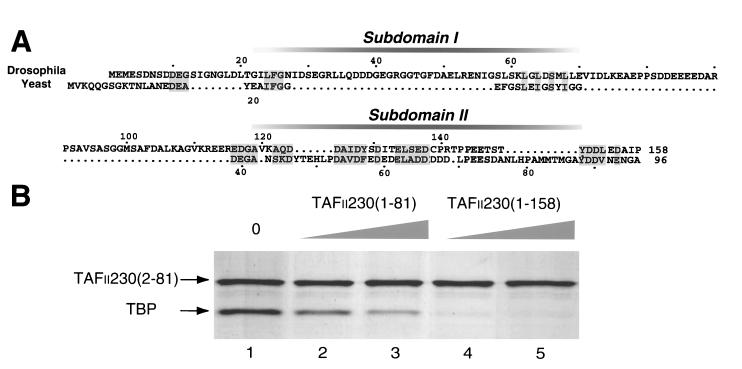
The transcription initiation factor TFIID, consisting of the TATA box-binding protein (TBP) and many TBP-associated factors (TAFs), plays a central role in both basal and activated transcription. An intriguing finding is that the 80-residue N-terminal region of Drosophila TAF(II)230 [dTAF(II)230-(2-81)] can bind directly to TBP and inhibit its function. Here, studies with mutated forms of TBP demonstrate that dTAF(II)230-(2-81) binds to the concave surface of TBP, which is important for TATA box binding. Previously, it was reported that a point mutation (L114K) on this concave surface destroys the ability of TBP to bind VP16 and to mediate VP16-dependent activation in vitro, but has no effect on basal transcription. Importantly, the same TBP mutation eliminates TBP binding to dTAF(II)230-(2-81). Consistent with these effects of the L114K mutation, dTAF(II)230-(2-81) and the VP16 activation domain compete for binding to wild-type TBP. These results indicate that transcriptional regulation may involve, in part, competitive interactions between transcriptional activators and TAFs on the TBP surface.


Figures

Figure 1
Figure 1
dTAFII230-(2–81) binds to the concave surface of TBP. (A) Ribbon drawing of TBP viewed perpendicular toward the internal pseudodyad axis and (B) space-filling drawing viewed to the concave surface. The binding capability of TBP mutants to dTAFII230-(2–81) is indicated by colors. Mutations that cause a weak or no effect are shown by yellow (>50% activity of wild-type TBP), whereas mutations that cause serious defects are shown by orange (2–10%) or red (<2%). (C) TBP interaction with dTAFII230-(2–81). GST–dTAFII230-(2–81) was incubated with wild-type (lanes 1–4) or mutant (lanes 5–18) TBP. GST–dTAFII230-(2–81) with alanine substitutions in five contiguous residues (residues 24–28) (ref. 17) was used as a control (lanes 3 and 4). Input (odd lanes) and purified (even lanes) materials were analyzed by SDS/PAGE and visualized by Coomassie brilliant blue staining. (D) Analysis of TBP–DNA interactions by gel shift assay. Reaction mixtures contained 0.5 (odd lanes) or 5 (even lanes) pmol of wild-type (lanes 1 and 2) or mutant (lanes 3–14) TBP. (E) Analysis of basal transcription activity. Transcription activity was determined in a TBP-dependent reconstituted transcription system with 1 pmol of wild-type (lane 1) or mutant (lanes 2–12) TBP. The position of the accurately initiated transcript is indicated by an arrow.
Figure 2
Figure 2
The VP16 activation domain inhibits TBP binding to the TATA box. All lanes except lane 1 contained 0.125 pmol of yTBP. The reaction mixture contained 12.5 pmol of GST (lane 3), GST–VP16 (lane 4), or GST–VP16Δ456 (lane 5).
Figure 3
Figure 3
dTAFII230-(1–81) and the VP16 activation domain competitively bind to TBP. GST (lane 1) or GST–VP16 (lanes 3–12) were incubated with 35S-labeled yTBP in the presence of histidine-tagged dTAFII230-(1–81) with alanine substitutions in five contiguous residues (residues 24-28) (ref. 17) (lanes 4–6), dTAFII230-(1–81) (lanes 7–9), or dTAFII230-(1–158) (lanes 10–12). Input (lane 1) and purified materials were analyzed by SDS/PAGE and visualized by autoradiography.
Figure 4
Figure 4
dTAFII230-(82–158) contributes to stable TBP binding. (A) Sequence alignment of N-terminal regions of dTAFII230 and yTAFII130. Dots indicate gaps introduced to maintain optimal alignment. Shading indicates residues identical or similar to each other. The positions of subdomains I and II are indicated. (B) TBP binding affinity of dTAFII230 N termini. GST–dTAFII230-(2–81) (40 pmol) was incubated with TBP (40 pmol) in the absence (lane 1) or in the presence of histidine-tagged dTAFII230-(1–81) (lanes 2 and 3) or histidine-tagged dTAFII230-(1–158) (lane 4 and 5) at 60 (lanes 2 and 4) and 200 (lanes 3 and 5) pmol. Binding experiments are represented as in Fig. 1C.

References

    1. Roeder R G. Trends Biochem Sci. 1996;21:327–335. - PubMed
    1. Koleske A J, Young R A. Trends Biochem Sci. 1995;20:113–116. - PubMed
    1. Halle J-P, Meisterernst Trends Genet. 1996;20:161–163. - PubMed
    1. Abmayr S M, Workman J L, Roeder R G. Genes Dev. 1988;2:542–553. - PubMed
    1. Horikoshi M, Carey M F, Kakidani H, Roeder R G. Cell. 1988;54:665–669. - PubMed

Publication types

LinkOut - more resources