Comparative Study
. 1997 Jan 7;94(1):91-6.
doi: 10.1073/pnas.94.1.91.

The motor domain and the regulatory domain of myosin solely dictate enzymatic activity and phosphorylation-dependent regulation, respectively

Affiliations
Comparative Study

The motor domain and the regulatory domain of myosin solely dictate enzymatic activity and phosphorylation-dependent regulation, respectively

M Sata et al. Proc Natl Acad Sci U S A. .

Abstract

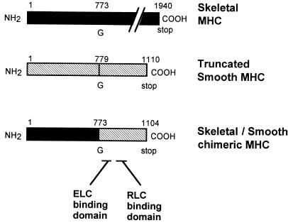
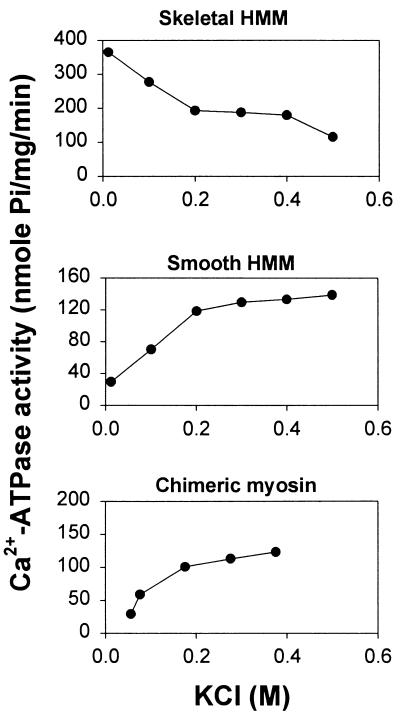
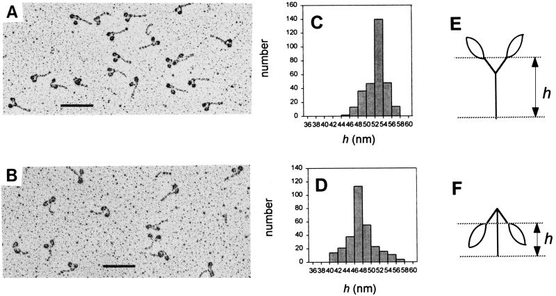
While the structures of skeletal and smooth muscle myosins are homologous, they differ functionally from each other in several respects, i.e., motor activities and regulation. To investigate the molecular basis for these differences, we have produced a skeletal/smooth chimeric myosin molecule and analyzed the motor activities and regulation of this myosin. The produced chimeric myosin is composed of the globular motor domain of skeletal muscle myosin (Met1-Gly773) and the C-terminal long alpha-helix domain of myosin subfragment 1 as well as myosin subfragment 2 (Gly773-Ser1104) and light chains of smooth muscle myosin. Both the actin-activated ATPase activity and the actin-translocating activity of the chimeric myosin were completely regulated by light chain phosphorylation. On the other hand, the maximum actin-activated ATPase activity of the chimeric myosin was the same as skeletal myosin and thus much higher than smooth myosin. These results show that the C-terminal light chain-associated domain of myosin head solely confers regulation by light chain phosphorylation, whereas the motor domain determines the rate of ATP hydrolysis. This is the first report, to our knowledge, that directly determines the function of the two structurally separated domains in myosin head.


Figures

Figure 1
Figure 1
Construction of skeletal/smooth chimeric MHC. Schematic drawing of skeletal/smooth chimeric MHC construct. A chimeric MHC cDNA was produced by ligating skeletal MHC cDNA containing the 5′ 2319 bp corresponding to the globular motor domain and a smooth MHC cDNA containing 993 bp corresponding to the C-terminal long α-helix domain of S1 and S2. ELC, essential light chain; RLC, regulatory light chain.
Figure 2
Figure 2
Isolation of the chimeric myosin.(A) SDS/PAGE (7.5–12% gel gradient) of myosin fragments. Lanes: 1, skeletal muscle myosin; 2, skeletal muscle HMM; 3, purified skeletal/smooth chimeric myosin; 4, smooth muscle myosin; 5, smooth muscle HMM; 6, molecular weight standard. (B) Immunoblot of the myosin fragments using a monoclonal antibody, 50KD, which recognizes a 50-kDa fragment of chicken skeletal muscle myosin S1. Lanes: 1, skeletal muscle myosin; 2, chimeric myosin; 3, smooth muscle myosin. (C) Immunoblot of the myosin fragments using a monoclonal antibody, MM9, which recognizes the S2 portion of chicken smooth muscle myosin. Lanes: 1, skeletal muscle myosin; 2, chimeric myosin; 3, smooth muscle myosin. (D) Nondenaturing gel electrophoresis of myosin fragments. Lanes: 1, smooth muscle HMM; 2, chimeric myosin; 3, smooth muscle myosin S1.
Figure 3
Figure 3
KCl dependence of Ca2+-ATPase activity of myosin fragments. Ca2+-ATPase activity was measured in 0.01 mg/ml myosin fragment, 5 mM CaCl2, 3 mM EDTA, 50 mM Tris·HCl (pH 7.5), and various concentrations of KCl at 25°C. The reaction was started by adding ATP, and the liberated inorganic phosphate was measured as described (20).
Figure 4
Figure 4
Apparent sedimentation coefficient distributions for myosin fragments. Sedimentation velocity was determined in solutions containing 2 mM MgCl2, 0.3 mM DTT, 1 mM ATP, 20 mM Tris·HCl (pH 7.5), 0.1 mg/ml myosin fragment, and either 0.01 M or 0.4 M KCl. The velocity runs were carried out at 56,000 rpm at 20°C for smooth HMM (A) and chimeric myosin (B). The x axis, formula image is the apparent sedimentation coefficient computed as described by Stafford (30, 31).
Figure 5
Figure 5
Electron micrographs of the chimeric myosin molecules. The rotary-shadowed images of the chimeric myosin in 0.4 M KCl (A) and 0.01 M KCl (B). (A and B, ×100,000; bars = 0.1 μm.) The distribution of the height of the globule/α-helix junction (h) in 0.4 M KCl and 0.01 M KCl are summarized in C and D, respectively. The height was measured according to the schematic drawings for the extended form (E) and the flexed form (F).

Similar articles

Cited by

References

    1. Mooseker M S, Cheney R E. Annu Rev Cell Dev Biol. 1995;11:633–675. - PubMed
    1. Zot A S, Potter J D. Annu Rev Biophys Biophys Chem. 1987;16:535–559. - PubMed
    1. Hartshorne D J. In: Physiology of the Gastrointestinal Tract. 2nd Ed. Johnson L R, editor. Vol. 1. New York: Raven; 1987. pp. 423–482.
    1. Sellers J R, Adelstein R A. In: The Enzymes. Boyer P, Krebs E G, editors. Vol. 18. San Diego: Academic; 1987. pp. 381–418.
    1. Trybus K M, Huiatt T M, Lowey S. Proc Natl Acad Sci USA. 1982;79:6151–6155. - PMC - PubMed

Publication types

MeSH terms

LinkOut - more resources