Comparative Study
. 1997 Jan 7;94(1):157-62.
doi: 10.1073/pnas.94.1.157.

Resistance of actin to cleavage during apoptosis

Affiliations
Comparative Study

Resistance of actin to cleavage during apoptosis

Q Song et al. Proc Natl Acad Sci U S A. .

Abstract

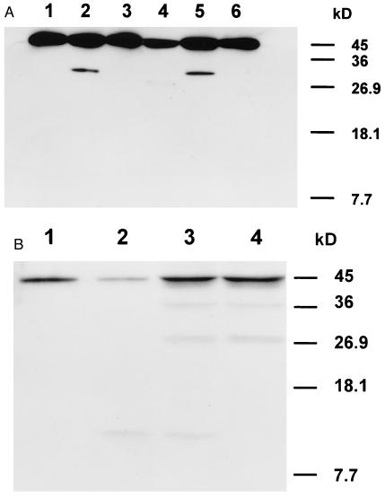
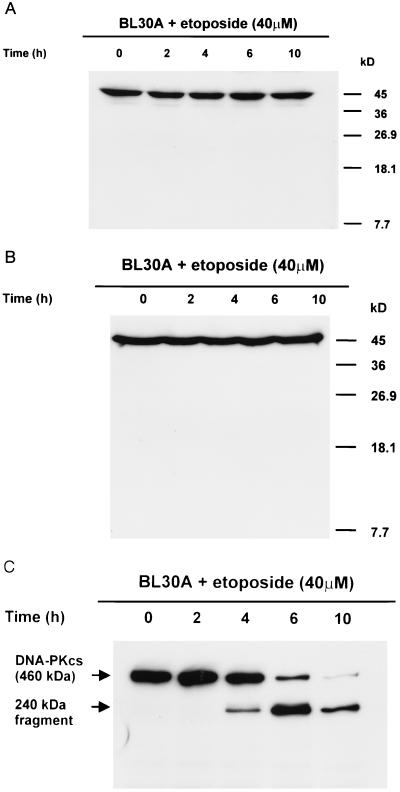
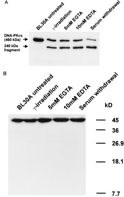
A small number of cellular proteins present in the nucleus, cytosol, and membrane fraction are specifically cleaved by the interleukin-1beta-converting enzyme (ICE)-like family of proteases during apoptosis. Previous results have demonstrated that one of these, the cytoskeletal protein actin, is degraded in rat PC12 pheochromocytoma cells upon serum withdrawal. Extracts from etoposide-treated U937 cells are also capable of cleaving actin. It was assumed that cleavage of actin represented a general phenomenon, and a mechanism coordinating proteolytic, endonucleolytic, and morphological aspects of apoptosis was proposed. We demonstrate here that actin is resistant to degradation in several different human cells induced to undergo apoptosis in response to a variety of stimuli, including Fas ligation, serum withdrawal, cytotoxic T-cell killing, and DNA damage. On the other hand, cell-free extracts from these cells and the ICE-like protease CPP32 were capable of cleaving actin in vitro. We conclude that while actin contains cleavage sites for ICE-like proteases, it is not degraded in vivo in human cells either because of lack of access of these proteases to actin or due to the presence of other factors that prevent degradation.


Figures

Figure 1
Figure 1
Cleavage of actin by ICE-like proteases and extracts from etoposide-treated BL30A cells. (A) Immunoblotting with a monoclonal antibody against the N terminus of actin, amino acids 23–34. Lane 1, actin alone; lane 2, CPP32 + actin; lane 3, TX + actin; lane 4, Mch2α + actin; lane 5, extracts from cells undergoing apoptosis + actin; and lane 6, extracts from cells undergoing apoptosis. (B) Immunoblotting with a polyclonal antibody against the C-terminal 11 amino acids of actin. Lane 1, actin alone; lane 2, CPP32 + actin; lane 3, extracts from cells undergoing apoptosis + actin; and lane 4, extracts from cells undergoing apoptosis.
Figure 2
Figure 2
Lack of cleavage of actin during apoptosis induced by etoposide in BL30A cells. (A) A polyclonal antibody against the C-terminal 11 amino acids of actin was used for the immunoblot. (B) Immunoblotting with a monoclonal antibody against amino acid region 23–34 of actin. (C) Time course of cleavage of DNA-PKcs after exposure of BL30A cells to 40 μM etoposide. DNA-PKcs was detected by using a polyclonal antibody against amino acid region 2018–2136 of DNA-PKcs (DPK1).
Figure 3
Figure 3
Lack of degradation of actin during apoptosis of BL30A cells induced by a variety of agents and serum withdrawal. (A) Effect of different agents and serum withdrawal on the cleavage of DNA-PKcs. Cells were treated with γ-irradiation (20 Gy) and incubated for 8 h; EGTA (5 mM) for 8 h; EDTA (10 mM) for 6 h; or serum was withdrawn for 24 h. In all cases, ≈40-60% of the cells were undergoing apoptosis. (B) Lack of degradation of actin after treatment of BL30A cells with the same agents and serum withdrawal. A polyclonal antibody against the C-terminal 11 amino acids of actin was used for the immunoblot.
Figure 4
Figure 4
Lack of actin cleavage in different cell types by a variety of agents causing apoptosis. (A) Lack of cleavage of actin in different Burkitt lymphoma cells after exposure of cells to 40 μM etoposide. A polyclonal antibody against the C-terminal 11 amino acids of actin was used for immunoblotting. (B) Cleavage of DNA-PKcs during apoptosis induced by etoposide (40 μM) in different Burkitt lymphoma cells. DNA-PKcs was detected with DPK1 antibody. (C) Lack of degradation of actin in HeLa, Molt-4, U937, and CTL cells undergoing apoptosis induced by different agents. For HeLa cells, extracts were prepared from detached cells after 8 h of treatment with etoposide (40 μM). For Molt-4 cells, cells were irradiated (20 Gy) and extracts were prepared 8 h after irradiation. For U937 cells, cells were pretreated with interferon γ (200 international units/ml) for 24 h and then incubated with anti-Fas antibody at 50 ng/ml for 10 h prior to preparation of extracts. CTLs were incubated with specific peptide for 4 h prior to preparation of extracts. Lane 1, Molt4 untreated; lane 2, Molt4 exposed to 20 Gy of γ-irradiation; lane 3, HeLa untreated; lane 4, HeLa + 40 μM etoposide; lane 5, CTL untreated; lane 6, CTL + specific peptide; lane 7, U937 untreated; lane 8, U937 + 50 ng/ml anti-Fas antibody. A polyclonal antibody against the C-terminal 11 amino acids of actin was used for immunoblotting. (D) Cleavage of DNA-PKcs during apoptosis induced by different agents in HeLa, Molt-4, U937, and CTL cells. DNA-PKcs was detected with DPK1 antibody.

Similar articles

Cited by

References

    1. Wyllie A H, Kerr J F R, Currie A R. Int Rev Cytol. 1980;68:251–306. - PubMed
    1. Steller H. Science. 1995;267:1445–1449. - PubMed
    1. White E. Genes Dev. 1996;10:1–15. - PubMed
    1. Vaux D L, Strasser A. Proc Natl Acad Sci USA. 1996;93:2239–2244. - PMC - PubMed
    1. Martin S J, Green D R. Cell. 1995;82:349–352. - PubMed

Publication types