. 1997 Jan 7;94(1):163-8.
doi: 10.1073/pnas.94.1.163.

A cellular 65-kDa protein recognizes the negative regulatory element of human papillomavirus late mRNA

Affiliations
Free PMC article

A cellular 65-kDa protein recognizes the negative regulatory element of human papillomavirus late mRNA

W Dietrich-Goetz et al. Proc Natl Acad Sci U S A. .
Free PMC article

Abstract

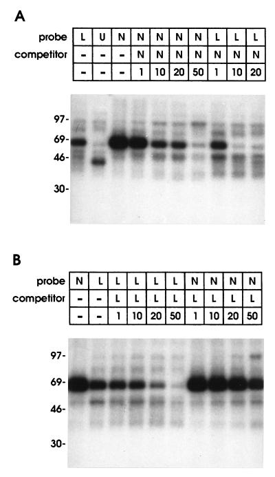
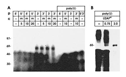
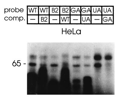
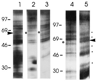
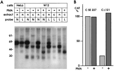
Papillomavirus late gene expression is tightly linked to the differentiation state of the host cell. Levels of late mRNAs are only in part controlled by regulation of the late promoter, other posttranscriptional mechanisms exist that reduce the amount of late mRNA in undifferentiated cells. Previously we described a negative regulatory element (NRE) located upstream of the human papillomavirus type 16 late poly(A) site. We have delineated the NRE to a 79-nt region in which a G+U-rich region was the major determinant of NRE activity. UV-crosslinking assays identified a prominent nuclear protein of 65 kDa as the only factor in close contact with the NRE, and a complex of at least five proteins, including the 65-kDa protein, was enriched on NRE-RNA. Binding of the 65-kDa protein was depleted by preincubation with poly(U) Sepharose in high salt, a property characteristic of the U2 small nuclear ribonucleoprotein auxiliary factor U2AF65 and bacterially expressed U2AF65 exhibited NRE binding. The 65-kDa protein bound to the G+U-rich NRE 3' half which shows homology to the B2P2 sequence a known U2AF65 binding site in the alpha-tropomyosin gene, and the G+U-rich element can be replaced by B2P2 in the binding assay. Treatment of cells with phorbol 12-myristate 13-acetate reduced binding of the 65-kDa protein, induced NRE binding of a cytoplasmic protein, and relieved the NRE block on reporter gene expression.


Figures

Figure 1
Figure 1
Boundaries of the HPV-16 NRE. The 5′ deletion series described earlier (6) was complemented by a 3′ deletion series. Effects on NRE activity were assayed using CAT reporter constructs with different NRE portions present upstream of the HPV-16 late poly(A) site. CAT activities relative to plasmid C-SE227 (100%) are indicated. ▴, 3′ deletion series; formula image
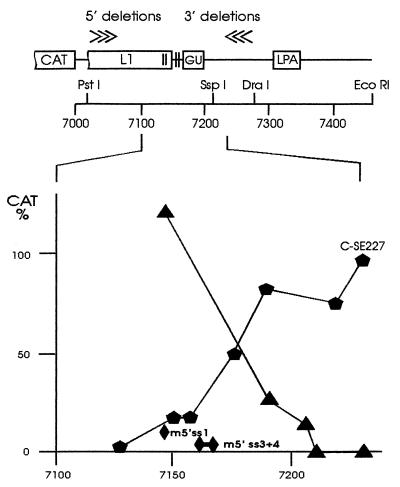
, 5′ deletion series; ♦, point mutations in 5′ splice site homologies 1 (m5′ss1) or 3 and 4 (m5′ss3+4).
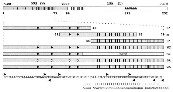
Figure 2
Figure 2
Templates used to generate radiolabeled riboprobes and unlabeled competitor RNAs. Probe N: 79-nt minimal NRE and 20 nt downstream (HPV-16 nt 7128–7226). Probe L: 153-nt region (nt 7227–7379) containing the HPV-16 late poly(A) site (LPA). Probe 5′: 5′ portion of the 79-nt NRE containing four 5′ splice site homologies (♦). | = uridines present in the 3′ portion of probe N and within the putative weak binding site in L. Probe m: middle NRE section containing two 5′ splice site homologies and half of the G+U-rich element. Probe 3′: 3′ NRE portion containing a G+U-rich region with homology to a B2P2 binding site. Probe WT: 79-nt wild-type NRE. Probe B2: G+U-rich region substituted by the U2AF65 binding site B2P2 from the α-tropomyosin gene. Probe GA: HPV-16 NRE with G to A mutations in the putative first intron base of all four 5′ splice site homologies ♦. Probe UA: six U to A mutations in the G+U-rich region (| |). Vertical bars in the L fragment indicate partial homology to the G+U-rich element. ▸, first nucleotides retained in the 5′ deletion clones; ◂, last nucleotides retained in the 3′ deletion clones. The B2P2 sequence is shown; vertical bars indicate nucleotide homologies, and colons refer to conserved purine/pyrimidine patterns.
Figure 3
Figure 3
Binding of HeLa cell nuclear proteins to the HPV-16 NRE. Nuclear proteins were UV-crosslinked to radiolabeled RNA probes spanning the NRE and 20 nt of downstream region (probe N), the adjacent 153-nt region (probe L), or the HSV-1 UL44 gene poly(A) site (probe U). A 1–50 molar excess of unlabeled probe was used for competition as indicated. (A) Competition of unlabeled riboprobe N against labeled probe N or labeled probe L. (B) Competition of unlabeled riboprobe L against labeled probe L or labeled probe N.
Figure 4
Figure 4
Binding properties of the 65-kDa protein and its target site within the NRE. (A) HeLa cell nuclear proteins were UV-crosslinked to probes corresponding to the 5′ half of the NRE (probe 5′) with four 5′ splice site homologies, or the 3′ half of the NRE (probe 3′) with a G+U-rich region with homology to a U2AF65 binding site. Poly(U) denotes nuclear extracts depleted by preincubation with poly(U) Sepharose in the presence of 2 M KCl. Unlabeled RNA corresponding to the middle section of the NRE (m, see Fig. 2) was used as competitor. (B) Reconstitution experiment with U2AF65. The indicated amounts (in micrograms) of partially purified, bacterial protein extract containing U2AF65 were added to HeLa cell nuclear extracts that were depleted of 65-kDa protein binding activity by preincubation with poly(U) Sepharose in 2 M KCl, probe N was used (Fig. 2).
Figure 5
Figure 5
Effects of mutations and substitutions in the NRE on 65-kDa protein binding. Wild-type and B2P2 substitutions (probes as in Fig. 2) were assayed for cross-competition with 10-fold molar excess of the respective unlabeled RNA, similarly the 5′ splice sites and G+U mutants were tested for cross-competition with their unlabeled counterparts with HeLa nuclear extract.
Figure 6
Figure 6
Purification of a larger NRE-associated protein complex. (A) Comparison of complete HeLa cell nuclear extract (lane 1) with extracts depleted by preincubation with biotinylated NRE–RNA and adsorption on streptavidin beads (lanes 2 and 4) or depleted by preincubation with poly(U) Sepharose in the presence of 2 M KCl (lane 3). Release of NRE-bound proteins from streptavidin beads by RNase treatment (lane 5). ▸, 65-kDa protein; ○, depleted 65-kDa protein; [cirf], enriched proteins; R, RNase A.
Figure 7
Figure 7
Effect of PMA treatment on NRE binding proteins and reporter gene expression. (A) Changes in protein binding of nuclear (n) or cytoplasmic (c) extracts from HeLa cells or W12 cells following treatment with PMA. (B) Changes of CAT activity in W12 cells transfected with CD121 or C-SE227 plasmids following treatment with PMA.

References

    1. Bedell M A, Hudson J B, Golub T R, Turyk M E, Hosken M, Wilbanks G D, Laimins L A. J Virol. 1991;65:2245–2260. - PMC - PubMed
    1. Meyers C, Frattini M G, Hudson J B, Laimins L A. Science. 1992;257:971–973. - PubMed
    1. Dollard S C, Wilson J L, Demeter L M, Bonney W, Reichman R C, Becker T R, Chow L T. Genes Dev. 1992;6:1131–1142. - PubMed
    1. Frattini M G, Lim H B, Laimins L A. Proc Natl Acad Sci USA. 1996;93:3062–3067. - PMC - PubMed
    1. Sterling J, Stanley M, Gatward G, Minson T. J Virol. 1990;64:6305–6307. - PMC - PubMed

Publication types

MeSH terms