

Penelope, a new family of transposable elements and its possible role in hybrid dysgenesis in Drosophila virilis
- PMID: 8990185
- PMCID: PMC19282
- DOI: 10.1073/pnas.94.1.196
Penelope, a new family of transposable elements and its possible role in hybrid dysgenesis in Drosophila virilis
Abstract
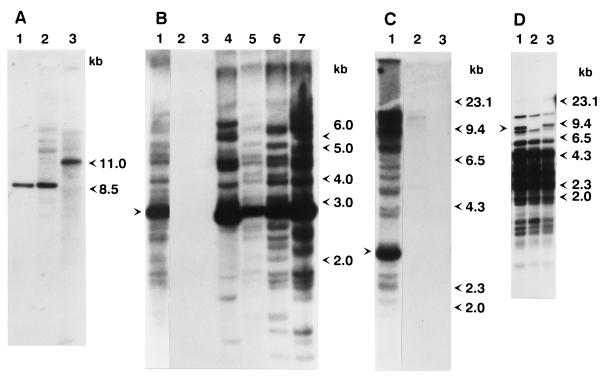
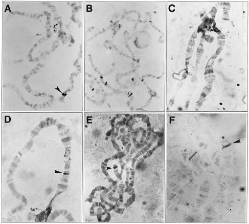
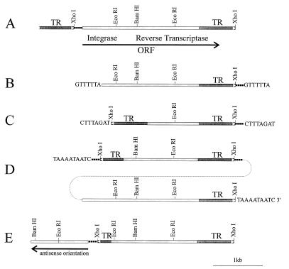
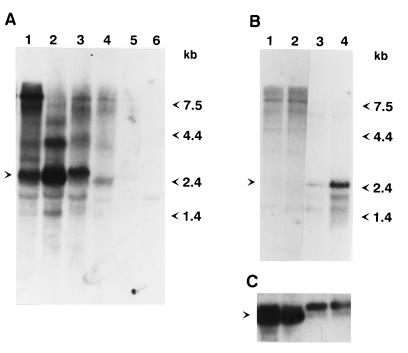
A hybrid dysgenesis syndrome occurs in Drosophila virilis when males from an established laboratory strain are crossed to females obtained from the wild, causing the simultaneous mobilization of several different transposable elements. The insertion sequence responsible for the mutant phenotype of a dysgenic yellow allele has been characterized and named Penelope. In situ hybridization and Southern analyses reveal the presence of more than 30 copies of this element in the P-like parental strain, whereas Penelope is absent in all M-like strains tested. Penelope contains one 2.5-kb-long ORF that could encode products with homology to integrase and reverse transcriptase. Northern analysis and whole-mount in situ hybridization show strong induction of a 2.6-kb RNA in the ovaries of dysgenic females that is expressed at very low levels in the parental strains or in the progeny from the reciprocal cross. Injection of Penelope-containing plasmids into preblastoderm embryos of an M-like strain results in mutant progeny caused by insertion of Ulysses and perhaps other transposons, suggesting that Penelope expression might be responsible for the observed dysgenesis syndrome and the simultaneous mobilization of other transposable elements.
Figures

Similar articles
-
[The unusual mobile element Penelope and its behavior in distant Drosophila species].Genetika. 2003 Feb;39(2):269-79. Genetika. 2003. PMID: 12669424 Russian.
-
The structure and evolution of Penelope in the virilis species group of Drosophila: an ancient lineage of retroelements.J Mol Evol. 2001 May;52(5):445-56. doi: 10.1007/s002390010174. J Mol Evol. 2001. PMID: 11443348
-
Expression of Drosophila virilis retroelements and role of small RNAs in their intrastrain transposition.PLoS One. 2011;6(7):e21883. doi: 10.1371/journal.pone.0021883. Epub 2011 Jul 11. PLoS One. 2011. PMID: 21779346 Free PMC article.
-
Penelope-like elements--a new class of retroelements: distribution, function and possible evolutionary significance.Cytogenet Genome Res. 2005;110(1-4):510-21. doi: 10.1159/000084984. Cytogenet Genome Res. 2005. PMID: 16093704 Review.
-
Genetic and molecular investigations on the endogenous mobile elements of non-drosophilid fruitflies.Genetica. 1997;100(1-3):119-29. Genetica. 1997. PMID: 9440264 Review.
Cited by
-
Paramutation-like Epigenetic Conversion by piRNA at the Telomere of Drosophila virilis.Biology (Basel). 2022 Oct 9;11(10):1480. doi: 10.3390/biology11101480. Biology (Basel). 2022. PMID: 36290385 Free PMC article.
-
Reverse transcriptase and endonuclease activities encoded by Penelope-like retroelements.Proc Natl Acad Sci U S A. 2004 Oct 12;101(41):14719-24. doi: 10.1073/pnas.0406281101. Epub 2004 Oct 1. Proc Natl Acad Sci U S A. 2004. PMID: 15465912 Free PMC article.
-
Novel ribozymes: discovery, catalytic mechanisms, and the quest to understand biological function.Nucleic Acids Res. 2019 Oct 10;47(18):9480-9494. doi: 10.1093/nar/gkz737. Nucleic Acids Res. 2019. PMID: 31504786 Free PMC article.
-
Extensive de Novo genomic variation in rice induced by introgression from wild rice (Zizania latifolia Griseb.).Genetics. 2005 Aug;170(4):1945-56. doi: 10.1534/genetics.105.040964. Epub 2005 Jun 3. Genetics. 2005. PMID: 15937131 Free PMC article.
-
Comparative polytene chromosome maps of D. montana and D. virilis.Chromosoma. 2007 Feb;116(1):21-7. doi: 10.1007/s00412-006-0075-3. Epub 2006 Aug 12. Chromosoma. 2007. PMID: 16906413
References
-
- Hiraizumi Y. Proc Natl Acad Sci USA. 1971;68:7369–7373.
-
- Sved J A. Aust J Biol Sci. 1976;29:375–388. - PubMed
-
- Kidwell M G, Kidwell J F, Sved J A. Genetics. 1977;86:333–351. - PubMed
-
- Rubin G M, Kidwell M G, Bingham P M. Cell. 1982;29:987–994. - PubMed
-
- O’Hare K, Rubin G M. Cell. 1983;34:25–35. - PubMed
Publication types
MeSH terms
Substances
Associated data
- Actions
Grants and funding
LinkOut - more resources
Full Text Sources
Other Literature Sources
Molecular Biology Databases
