

doi: 10.1073/pnas.94.1.213.
Beta cell apoptosis in T cell-mediated autoimmune diabetes
Affiliations
- PMID: 8990188
- PMCID: PMC19288
- DOI: 10.1073/pnas.94.1.213
Item in Clipboard
Beta cell apoptosis in T cell-mediated autoimmune diabetes
Proc Natl Acad Sci U S A.
.
Abstract
Insulin-dependent diabetes mellitus results from T cell-mediated destruction of insulin-producing, pancreatic islet beta cells. How this destruction takes place has remained elusive--largely due to the slow kinetics of disease progression. By crossing a transgenic mouse carrying a beta cell-specific T cell receptor onto the NOD.scid background, we produced a simplified but robust and accelerated model of diabetes. This mouse produces CD4+ T cells bearing transgenic T cell receptor but is devoid of CD8+ T cells and B cells. More importantly, this mouse develops a rapid diabetes, which has allowed us to record and quantify beta cell death. We have determined that beta cells within the inflamed islets die by apoptosis.
Figures

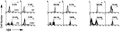
Transgenic TCR expression selects CD4+ T cells in the thymi of BDC2.5/NOD.scid mice. Three-color flow cytometric analysis of thymi from negative littermate (NOD.scid/+; Left), TCR transgenic (BDC2.5/NOD.scid/+; Center), and TCR/NOD.scid transgenic (BDC2.5/NOD.scid; Right) mice was performed using anti-CD4, anti-CD8α, and anti-Vβ4 TCR mAb. (Upper) Two-color dot plots of CD4 versus CD8. (Lower) Vβ4 TCR expression on gated CD4+CD8− (CD4+), CD4+CD8+ (DP), CD4−CD8+ (CD8+) and CD4− CD8− (DN) subpopulations.
Transgenic TCR expression selects CD4+ T cells in the thymi of BDC2.5/NOD.scid mice. Three-color flow cytometric analysis of thymi from negative littermate (NOD.scid/+; Left), TCR transgenic (BDC2.5/NOD.scid/+; Center), and TCR/NOD.scid transgenic (BDC2.5/NOD.scid; Right) mice was performed using anti-CD4, anti-CD8α, and anti-Vβ4 TCR mAb. (Upper) Two-color dot plots of CD4 versus CD8. (Lower) Vβ4 TCR expression on gated CD4+CD8− (CD4+), CD4+CD8+ (DP), CD4−CD8+ (CD8+) and CD4− CD8− (DN) subpopulations.

Lack of CD8+ T cells in the spleen of BDC2.5/NOD.scid mice. Three-color flow cytometric analysis of splenocytes from negative littermate (NOD.scid/+; Left), TCR transgenic (BDC2.5/NOD.scid/+; Center), and TCR/NOD.scid (BDC2.5/NOD.scid; Right) mice was performed using anti-CD4, anti-CD8α, and anti-Vβ4 TCR mAb. (Top) Two-color CD4 versus CD8 dot plots. (Middle) Vβ4 TCR expression on gated CD4+CD8− (CD4+), CD4−CD8+ (CD8+) and CD4−CD8− (DN) subpopulations. (Bottom) Two-color dot plots, B220 versus sIgM confirms scid phenotype of B cells in bottom right panel.

Rapid insulitis and diabetes in BDC2.5/NOD.scid mice. (a) Insulitis time course in BDC2.5/NOD.scid (filled bars) and BDC2.5/NOD.scid/+ littermates (open bars). Multiple hematoxylin and eosin pancreas sections from 3–6 mice in each time group were analyzed for insulitis. Eighty to 100 islets from each mouse were scored for no insulitis (0), peri- and mild insulitis (1), moderate insulitis (2), or severe insulitis (3), and then each mouse was given a mean score of insulitis. (b) The kinetics of diabetes was determined by measurement of blood glucose using a glucometer. Animals were considered diabetic after two consecutive readings ≥250 mg/dl (13.75 mM). •, BDC2.5/NOD.scid mice; ○, BDC2.5/NOD.scid/+ littermates. No diabetic BDC2.5/NOD.scid mouse lived past 33 days of age unless treated with insulin.
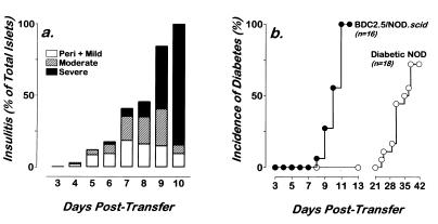
T cells from diabetic BDC2.5/NOD.scid mice efficiently transfer insulitis and diabetes to NOD.scid recipients. NOD.scid mice received 106 T cells from BDC2.5/NOD.scid mice by intravenous injection. Recipients were then followed for evidence of insulitis and diabetes. (a) Insulitis was assessed from multiple hematoxylin and eosin sections of pancreas. Insulitis was scored as peri- and mild insulitis (open bars), moderate (hatched bars), or severe (filled bars). Insulitis is represented as the composite of 40–100 islets per animal and 3–6 mice per time point. (b) Cumulative incidence of diabetes in 16 NOD.scid recipients of 106 T cells from diabetic BDC2.5/NOD.scid mice. Diabetes was assessed by measurement of blood glucose using a glucometer. Mice were considered diabetic after two consecutive readings ≥250 mg/dl (13.75 mM). Diabetes was dated to the first sequential readings.
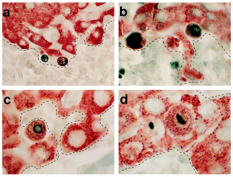
β cell apoptosis in BDC2.5/NOD.scid mice. Photomicrographs of TUNEL+ β cells at site of leukocytic infiltration. TUNEL+ nuclei stained with DAB of β cells (insulin containing granule stained with AEC) at the interface of leukocytic infiltration. The dashed lines demarcate the interface of insulitis as determined from parallel hematoxylin-stained sections. (a) ×250 magnification; (b) ×400 magnification; and (c and d) ×500 magnification.
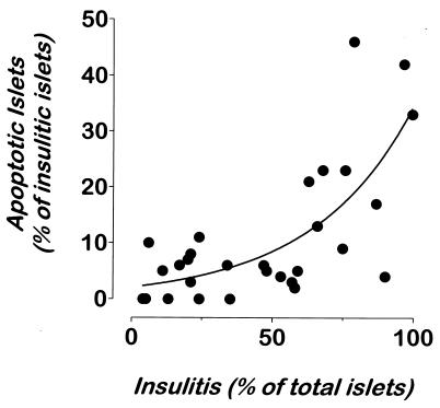
Presence of apoptotic β cells correlates with the level of insulitis. Pancreata from 31 normoglycemic NOD.scid recipients of T cells from diabetic BDC2.5/NOD.scid mice were analyzed for insulitis and apoptosis. Multiple sections from five distinct layers from each pancreas were stained by TUNEL and anti-insulin reagents and with hematoxylin. Islets were scored independently for insulitis and apoptosis. On average, 100 islets were scored per mouse. A second-order relationship (solid curve) was found using the graphpad software (r = 0.8963).
References
Publication types
MeSH terms
Substances
Grants and funding
LinkOut - more resources
Full Text Sources
Medical
Research Materials
