. 1997 Jan 7;94(1):219-26.
doi: 10.1073/pnas.94.1.219.

Transgenic studies with a keratin promoter-driven growth hormone transgene: prospects for gene therapy

Affiliations

Transgenic studies with a keratin promoter-driven growth hormone transgene: prospects for gene therapy

X Wang et al. Proc Natl Acad Sci U S A. .

Abstract

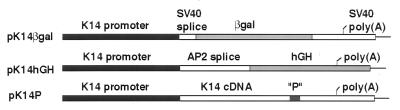
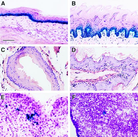
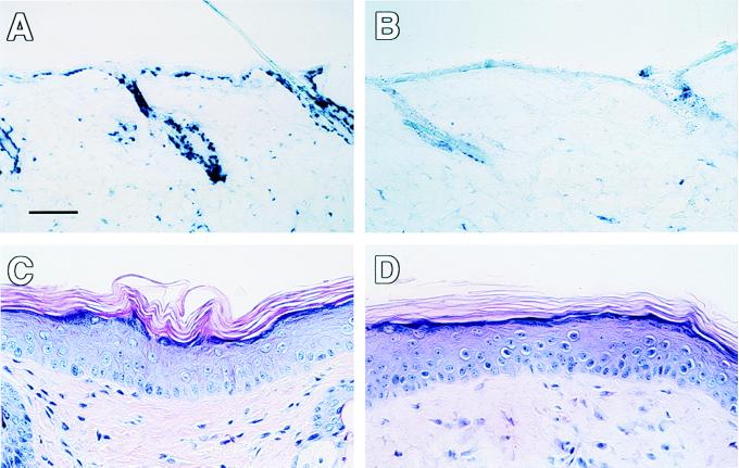
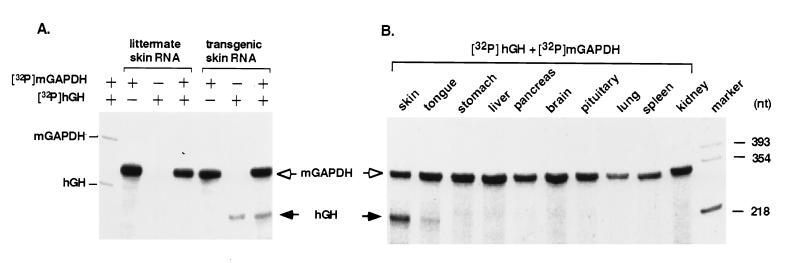
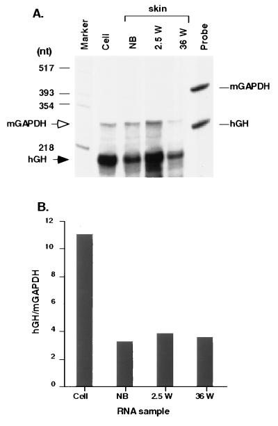
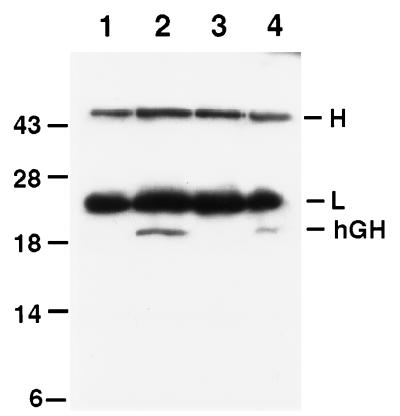
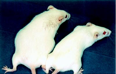
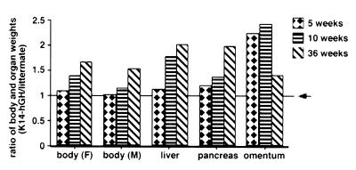
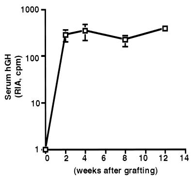
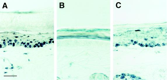
Keratinocytes are potentially appealing vehicles for the delivery of secreted gene products because they can be transferred to human skin by the relatively simple procedure of grafting. Adult human keratinocytes can be efficiently propagated in culture with sufficient proliferative capacity to produce enough epidermis to cover the body surface of an average adult. However, the feasibility of delivering secreted proteins through skin grafting rests upon (i) the strength of the promoter in keratinocytes and (ii) the efficiency of protein transport through the basement membrane of the stratified epithelium and into the bloodstream. In this paper, we use transgenic technology to demonstrate that the activity of the human keratin 14 promoter remains high in adult skin and that keratinocyte-derived human growth hormone (hGH) can be produced, secreted, and transported to the bloodstream of mice with efficiency that is sufficient to exceed by an order of magnitude the circulating hGH concentration in growing children. Transgenic skin grafts from these adults continue to produce and secrete hGH stably, at approximately 1/10 physiological levels in the bloodstream of nontransgenic recipient mice. These studies underscore the utility of the keratin 14 promoter for expressing foreign transgenes in keratinocytes and demonstrate that keratinocytes can be used as effective vehicles for transporting factors to the bloodstream and for eliciting metabolic changes. These findings have important implications for considering the keratinocyte as a possible vehicle for gene therapy.


Figures

Figure 1
Figure 1
Expression constructs used for the generation of transgenic mice. Stick diagrams depict the K14 promoter constructs engineered to drive expression of transgenes. Plasmid pK14βgal contains the 2.1-kb AvaI fragment containing the K14 promoter/enhancer, extending to the transcription initiation site. This fragment was followed by the intron present in the 5′-untranslated sequence of the simian virus 40 (SV40) large T antigen gene (SV40 splice), followed by a BamHI site in which the β-gal gene was inserted, followed by the 3′-untranslated sequences and polyadenylylation site from the SV40 large T antigen cDNA (SV40 polyA). Plasmid pK14hGH is similar in design and yields an indistinguishable expression pattern from pKβgal, but contains 1.1 kb of sequences from the AP2 gene (AP2 splice) in its BamHI site (these sequences did not interfere with hGH expression and are irrelevant for the purposes outlined in this paper). The entire 2.1-kb hGH gene followed, including introns, 3′-untranslated sequence, and polyadenylylation signal. An additional reporter construct was used, previously referred to as pH3cK14 promoter (16), and referred to here as pK14 promoter. Plasmid K14 promoter contained the K14 promoter, followed by the complete K14 coding sequence, modified at its 3′ end to add a small epitope tag sequence encoding the antigenic portion of neuropeptide substance P, followed by 3′-untranslated sequences and polyadenylylation signal of the K14 gene (16).
Figure 2
Figure 2
β-Gal activity assays in tissue sections from pK14βgal transgenic mice. Transgenic mice positive for the pK14βgal transgene were generated as described. Tissues were collected and processed for β-gal activity assays and for hematoxylin/eosin counterstaining as described. Most of the data on transgene expression are compiled in Table 1. Shown are representative examples of sections from tailskin (A), dorsal portion of tongue (B), esophagus (C), forestomach (D), salivary gland (E), and thymus (F). Note that for esophagus and forestomach, samples were also examined without counterstaining to verify that no β-gal activity was detected (not shown). Note also that only a few myoepithelial cells of the salivary gland and even fewer reticular cells of the thymic epithelium stained positive for the transgene. (Bar = 67 μm in AD and F, and 42 μm in E.)
Figure 3
Figure 3
Ectopic expression of the hGH transgene in skin does not alter epidermal morphology. (A and B) Backskin sections from an adult line 22 K14–hGH expressing mouse (A) and a control littermate (B) were subjected to in situ hybridization with an antisense digoxygenin-labeled hGH cRNA. (C and D) Tailskin sections from an adult transgenic (C) and control (D) mouse were stained with hematoxylin/eosin. Note that the epidermis of backskin, with abundant hair follicles, is thinner than that of tailskin, with few follicles. (Bar = 90 μm in A and B, and 45 μm in C and D.)
Figure 4
Figure 4
RNase protection assays to quantitate hGH RNA levels in transgenic mouse tissues. RNase protection assays were carried out as described. (A) Assays on adult skin. First lane shows migration of aliquot of antisense RNAs before RNase digestion. Other lanes show migration of radiolabeled antisense probes after incubation with skin RNAs and RNase treatment. Note the reduction in size for both mGAPDH (open arrowhead at right) and hGH (filled arrowhead at right) RNAs, reflective of digestion of the random (i.e., nonhybridizing) sequences in each of the two probes. This serves as an internal control for the RNase. (B) Assays on tissues. Lanes show protected radiolabeled cRNAs after hybridization with 9-month-old transgenic tissue RNAs and subsequent RNase treatment. Last lane shows migration of HinfI-digested pSP64 plasmid DNAs, end-labeled and used as molecular mass markers. (Note, the specific activity of the mGAPDH probe was higher in A versus B.)
Figure 5
Figure 5
RNase protection assays to quantitate hGH RNA levels in transgenic mouse skin at different ages. (A) RNAs were isolated from transgenic skin at birth (newborn, NB), and at 2.5 and 36 weeks postnatally. RNAs were also isolated from cultured transgenic mouse keratinocytes (Cell). RNase protection assays were carried out as described in the legend to Fig. 3, except that the specific activity of the radiolabeled mGAPDH antisense probe added to each sample was less than that used previously. Following hybridization and RNase treatment, the protected RNA fragments were resolved by electrophoresis through 6% acrylamide gels containing 8 M urea. (B) Phosphoimage analysis of blot in A. The amount of hybridizing radioactivity in each band was determined to obtain the ratio of protected hGH RNA relative to internal mGAPDH RNA in each sample. Relative to the level of mGAPDH RNAs in skin, the level of hGH transgene RNA did not vary significantly over time.
Figure 6
Figure 6
Immunoblot analysis of anti-hGH immunoprecipitates from sera of hGH transgenic mice and from the spent medium of keratinocytes cultured from newborn animals. Sera were taken from the bloodstream of 9-month-old transgenic and control mice. Keratinocytes from newborn control and transgenic mice were cultured in duplicate as described; at 80% confluence, fresh medium added, and the medium was sampled after an additional 24 hr of culture. Immunoprecipitations to detect the hGH transgene product in the skin or in the culture medium were carried out using mAb-coated plastic beads for hGH (Hybritech) under the same conditions as RIAs (see Materials and Methods), but without radiolabeled tracer. Aliquots of the immunoprecipitates were then resolved by SDS/PAGE and subjected to immunoblot assays, performed with a mAb against hGH (Sigma). Immunoprecipitates were from control serum (lane 1), transgenic serum (lane 2), control medium (lane 3), and transgenic medium (lane 4). Migration of molecular mass standards are indicated at left in kilodaltons; migration of heavy (H) and light (L) chains of γ-immunoglobulin is given at right.
Figure 7
Figure 7
K14–hGH transgenic mice are larger than control animals. Shown are a 2-month-old K14–hGH transgenic mouse (Left) and its control littermate (Right) from line 22. Note the increase in body size of the hGH-expressing mouse.
Figure 8
Figure 8
Weights of K14–hGH transgenic and control animals and organs. Male K14–hGH transgenic and control littermates were killed and/or weighed at the ages indicated. Organs were removed and weighed from 3–5 animals in each group, and an average weight was calculated. Shown is a plot of the ratio of transgenic versus control body and organ weights for each age group. Arrow denotes control level (ratio = 1). Because transgenic line 22 and 39 did not vary significantly in their organ weights, the values shown represent a combination of the measurements on the two lines. Shown for comparison are the data from MtI–hGH transgenic mice reported previously by Palmiter et al. (32, 40).
Figure 9
Figure 9
Detection of hGH in the bloodstream of SCID mice grafted with a patch of K14–hGH tailskin. The tailskins of 2-month-old K14–hGH transgenic mice were removed and grafted in 1–2 cm2 segments onto the backs of six recipient immunodeficient SCID mice. Animals were allowed to recover for 1–2 weeks, after which time blood was analyzed by RIA for hGH. To collect sufficient blood at 1-week intervals, samples from two animals were often combined for the assay, yielding three measurements from each week. The variation in levels are indicated by the vertical bars. To date, animals have been monitored for 14 weeks after the graft. After an initial recovery period after grafting, the concentrations of serum hGH were consistently within the range of 0.1–0.4 ng/ml.
Figure 10
Figure 10
In situ hybridization revealing hGH mRNA in the epidermis of K14–hGH skin grafts. A skin biopsy from one of the tailskin grafts of a SCID mouse recipient was removed and processed for in situ hybridization with a digoxygenin-labeled antisense hGH cRNA probe. (A) Biopsy from tailskin graft. (B) Control tailskin. (C) Tailskin from a K14–hGH transgenic mouse. Note that the dead, s. corneum layer occasionally traps some labeling (see example in B), but this is an artifact and is not consistently observed. Note the complete absence of labeling in the living epidermal layers of control skin (B). Note that the cells in the dermis of A and C that show labeling are from segments of hair follicles. (Bar = 30 μm.)

References

    1. Rochat A, Kobayashi K, Barrandon Y. Cell. 1994;76:1063–1073. - PubMed
    1. Rheinwald J G, Green H. Cell. 1975;6:331–343. - PubMed
    1. Rheinwald J G, Green H. Nature (London) 1977;265:421–424. - PubMed
    1. Gallico G G, O’Connor N E, Compton C C, Kehinde O, Green H. N Eng J Med. 1984;311:448–451. - PubMed
    1. Morgan J R, Barrandon Y, Green H, Mulligan R C. Science. 1987;237:1476–1479. - PubMed

Publication types

MeSH terms