. 1997 Jan 7;94(1):283-6.
doi: 10.1073/pnas.94.1.283.

The choroid plexus epithelium is the site of the organic anion transport protein in the brain

Affiliations

The choroid plexus epithelium is the site of the organic anion transport protein in the brain

R H Angeletti et al. Proc Natl Acad Sci U S A. .

Abstract

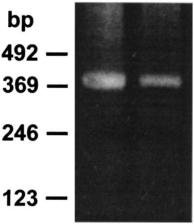
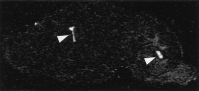
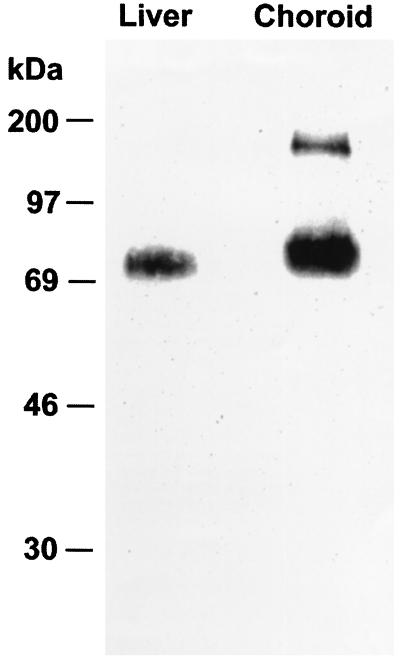
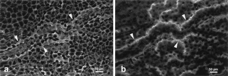
The mRNA for organic anion transport protein (oatp) was previously shown to be present in abundance in liver and kidney, and in small amounts in brain. Data obtained from experiments with reverse transcriptase-PCR techniques and in situ hybridization analysis showed that the oatp mRNA is present within the brain, localized to the choroid plexus. A sequence-specific antibody to the oatp polypeptide demonstrated the presence of the expected polypeptide with a molecular weight of 80,000 plus an immunoreactive species with a higher molecular weight in preparations of choroid plexus membranes. Examination of the choroid plexus by fluorescence confocal microscopy revealed that immunoreactive oatp polypeptide is localized to the apical surface of the choroid plexus epithelial cells, which contacts the cerebrospinal fluid. This localization of oatp is consistent with previous experiments showing vectorial transport of organic anions between the choroid plexus and the cerebrospinal fluid.


Figures

Figure 1
Figure 1
RT-PCR analysis of oatp mRNA in choroid plexus. The reaction products separated by agarose gel electrophoresis are shown for mRNA prepared from liver (left lane) and choroid plexus (right lane). The molecular weight markers are shown on the left.
Figure 2
Figure 2
In situ hybridization analysis of oatp mRNA in rat brain. A sagittal section of the brain is shown after hybridization with a radiolabeled, oatp-specific oligonucleotide probe and exposure to film. Arrowheads point out the intense labeling in the lateral (left arrowhead) and fourth (right arrowhead) ventricles.
Figure 3
Figure 3
Immunoblot analysis of the oatp polypeptide. The immunoreactive bands in liver (left lane) and choroid plexus (right lane) are shown. Molecular weight markers are at the left.
Figure 4
Figure 4
Confocal microscopy of choroid plexus. Freshly extirpated choroid plexus was incubated either with the fluorescein isothiocyanate derivative of phalloidin to reveal actin (a) or antiserum to oatp followed by a rhodamine-labeled secondary antibody (b). Arrowheads point out the intense staining seen at the apical surface of the epithelial cells.

References

    1. Pappenheimer J R, Heisey S R, Jordan E F. Am J Physiol. 1961;200:1–10. - PubMed
    1. Cameron G. Anat Rec. 1953;117:115–123. - PubMed
    1. Barany E H. Acta Physiol Scand. 1973;88:412–429. - PubMed
    1. Bratlid D. Clin Perinatol. 1990;17:449–465. - PubMed
    1. Jacquemin E, Hagenbuch B, Stieger B, Wolkoff A W, Meier P J. Proc Natl Acad Sci USA. 1994;91:133–137. - PMC - PubMed

Publication types

MeSH terms

LinkOut - more resources