Review
. 1997 Jan 7;94(1):343-50.
doi: 10.1073/pnas.94.1.343.

Place cells and place navigation

Affiliations
Review

Place cells and place navigation

J Bures et al. Proc Natl Acad Sci U S A. .

Abstract

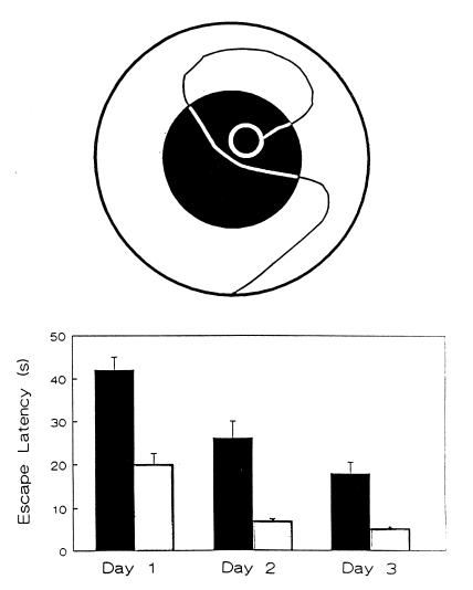
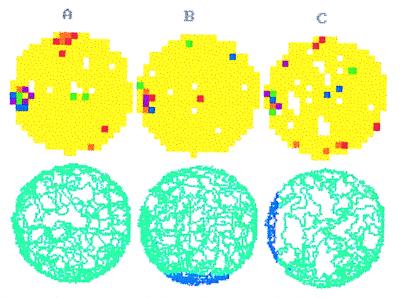
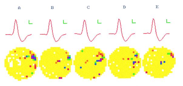
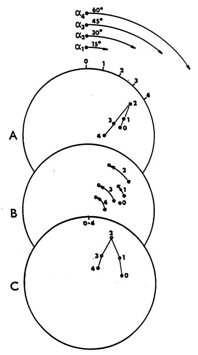
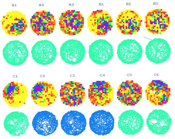
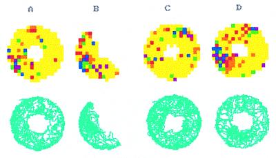
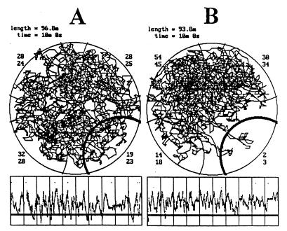
The assumption that hippocampal place cells (PCs) form the neural substrate of cognitive maps can be experimentally tested by comparing the effect of experimental interventions on PC activity and place navigation. Conditions that interfere with place navigation (darkness, cholinergic blockade) but leave PC activity unaffected obviously disrupt spatial memory at a post-PC level. Situations creating a conflict between egocentric and allocentric orientation (place navigation in the Morris water maze filled with slowly rotating water) slow down spatial learning. PC recording in rats searching food pellets in a rotating arena makes it possible to determine which firing fields are stable relative to the room (allocentrically dependent on sighted extramaze landmarks), to the surface of the arena (dependent on egocentric path integration mechanisms and intra-arena cues), or disappear during rotation. Such comparison is made possible by the computerized tracking system simultaneously displaying a rat's locomotion and the respective firing rate maps both in the room reference and arena reference frames. More severe conflict between allocentric and egocentric inputs is produced in the field clamp situation when the rat searching food in a ring-shaped arena is always returned by rotation of the arena to the same allocentric position. Ten-minute exposure to this condition caused subsequent disintegration or remapping of 70% PCs (n = 100). Simultaneous examination of PC activity and navigation is possible in the place avoidance task. A rat searching food in a stationary or rotating arena learns to avoid an allocentrically or egocentrically defined location where it receives mild electric footshock. In the place preference task the rat releases pellet delivery by entering an unmarked goal area and staying in it for a criterion time. Both tasks allow direct comparison of the spatial reference frames used by the PCs and by the behaving animal.


Figures

Figure 1
Figure 1
Impaired navigation to the escape platform located in the zero-visibility zone. (Upper) The Inset shows the scheme of the experiment with the zero-visibility zone marked by the black circle and the escape platform by the white ring in the northeast quadrant of the pool. The rat is started from the south and the room lights are on (black track) or off (white track) when it is outside or inside the zero-visibility zone, respectively. (Lower) Mean (± SEM) escape latencies of naive rats learning the task in light (n = 10, white columns) and with the goal in the zero-visibility zone (n = 10, black columns) during the first 3 days of training (12 trials/day). [This figure was modified from Arolfo et al. (51).]
Figure 2
Figure 2
Spatial firing characteristics of a hippocampal PC in a rat performing the pellet chasing task in a uniformly illuminated arena (2 m in diameter) before (A) and after introduction of the zero visibility are zones in the south (B) or west (C). (Lower) The tracks of the animal during 10-min recording sessions. The zero-visibility zones are indicated by dark tracking lines. The median firing rates from yellow to purple are 0.0, 0.2, 0.4, 1.0, 1.3, and 6.0 for A; 0.0, 0.1, 0.3, 0.4, 0.5, and 0.6 for B; and 0.0, 0.1, 0.4, 0.6, 0.8, and 2.9 for C. (See text for a quantitative evaluation of the maps.)
Figure 3
Figure 3
Activity of a hippocampal PC during pellet chasing in the arena used in Fig. 2 before (A) and during four successive 10-min-long recording periods (BE) after intraperitoneal injection of scopolamine (1 mg/kg). (Upper) The averaged shape of the selected spike (calibration 60 μV, 0.1 ms). (Lower) The firing rate maps. The median firing rates for the color coded pixels are 0.0, 0.2, 0.5, 1.0, 1.2, and 2.3 for A; 0.0, 0.3, 0.6, 0.9, 1.1, and 1.8 for B; 0.0, 0.1, 0.5, 0.8, 1.3, and 2.0 for C; 0.0, 0.3, 0.5, 1.4, 1.9, and 3.3 for D; 0.0, 0.1, 0.2, 0.3, 0.8, and 1.4 for E. (For quantitative evaluations, see text.)
Figure 4
Figure 4
Scheme of the transformation of the room frame display as seen by the real overhead camera (A) to the arena frame display as seen by a virtual overhead camera attached to the arena (C). The rat’s room-related locomotion on an arena rotating at constant angular velocity (15°/s) is transformed to arena-centered coordinates. During transformation (B) each position of the rat seen in A (points 0–4) remains at the same radial distance from the center of the arena but its angular coordinate is corrected by the angle corresponding to the angular displacement of the arena indicated by the position of the simultaneously recorded LED marker at its periphery (short radial bars 0 to 4 indicating the position of the LED at time intervals 0–4 s in A).
Figure 5
Figure 5
Firing rate maps of hippocampal PCs in a small (1 m in diameter) stationary arena and steadily rotating (one revolution per min) arena recorded in the room reference and arena reference projections. Other description as in Fig. 2 and 3. (A) A FF in the southeast quadrant of the stationary arena (A1) disappears during rotation both in the room reference (A2) and arena reference (A3) projections, while the firing rate of the PC increase 4 times. (B) Another FF in the southeast quadrant of the stationary arena (B1) is partly preserved during rotation of the arena in the room reference (B2) but not in the arena reference (B3) projections. (C) Another FF in the northwest quadrant of the stationary arena (C1) remains preserved in darkness (C2). During rotation in darkness the FF disappears in the room frame (C3) but not in the arena frame (C4). Similar effect of rotation was also observed in light provided that a cue card was placed on the arena wall: the FF disappeared in the room frame (C5) but was preserved in the arena frame (C6) albeit slightly shifted clockwise. The median firing rates for the color coded pixels are 0.0, 0.5, 1.1, 2.0, 2.9, and 4.0 for A1; 0.0, 0.8, 1.7, 2.5, 3.7, and 6.0 for A2; 0.0, 0.9, 1,6, 2.5, 3.7, and 5.0 for A3; 0.0, 0.6, 1.3, 2.6, 3.9, and 6.2 for B1; 0.0, 0.3, 0.8, 1.7, 2.7, and 5.0 for B2; 0.0, 0.4, 0.8, 1.2, 2.2, and 4.3 for B3; 0.0, 0.5, 1.2, 2.7, 5.4, and 9.0 for C1; 0.0, 0.6, 1.1, 2.5, 5.2, and 8.0 for C2; 0.0, 0.6, 1.2, 1.9, 2.9, and 5.0 for C3; 0.0, 0.6, 1.2, 2.2, 3.1, and 6.0 for C4; 0.0, 0.9, 1.8, 2.5, 3.5, and 5.0 for C5; 0.0, 0.7, 1.7, 2.9, 4.1, and 5.9 for C6. (For quantitative evaluations, see the text.)
Figure 6
Figure 6
Firing rate maps of a hippocampal PC recorded during pellet chasing in the ring-shaped arena before (A), during (B), immediately after (C) and 1 h after (D) exposure to the field clamp situation. In B rotation of the arena always returns the rat to a 30° segment between 150° and 180°. Other description as in Fig. 2. The median firing rates for the color coded pixels are 0.0, 0.2, 0.4, 0.5, 0.8, and 1.5 for A; 0.0, 0.2, 0.3, 0.6, 1.1, and 1.4 for B; 0.0, 0.2, 0.3, 0.4, 0.8, and 1.2 for C; 0.0, 0.2, 0.6, 1.2, 1.7, and 3.0 for D. (For quantitative evaluations, see the text.)
Figure 7
Figure 7
Tracking of a rat searching pellets on a slowly moving (one revolution per 30 s) circular arena (1 m diameter) before (A) and during (B) acquisition of the place avoidance induced by electric foot shocks applied whenever the animal enters a southeast region of the floor (marked by the heavy semicircular line). Length, total length of the track; time, total recording time. The numbers in the corners indicate the percentage of track length and track time corresponding to four equal semicircular regions in northeast, southeast, southwest, and northwest. The lower curves show the rat’s distance from the center of the shock region during the 10-min recording, with the critical radius indicated by the heavy horizontal line. The vertical lines indicate 1-min intervals. Note that all regions were equally visited before electrical stimulation in A and that the shock region was avoided after the two shocks received in the first and second minute of exploration in B.

Similar articles

Cited by

References

    1. Leao A A P. J Neurophysiol. 1944;7:359–390.
    1. Buresova O. Physiol Bohemoslov. 1956;5:350–358. - PubMed
    1. Bures J, Buresova O, Krivanek J. The Mechanism and Applications of Leao’s Spreading Depression of EEG Activity. New York: Academic; 1974.
    1. Bures J, Buresova O, Huston J P. Techniques and Basic Experiments for the Study of Brain and Behavior. Amsterdam: Elsevier; 1976.
    1. Bures J, Buresova O, Krivanek J. Brain and Behavior: Paradigms for Research in Neural Mechanisms. Chichester, U.K.: Wiley; 1988.

Publication types

LinkOut - more resources