. 1997 Feb 1;17(3):875-81.
doi: 10.1523/JNEUROSCI.17-03-00875.1997.

The Drosophila erg K+ channel polypeptide is encoded by the seizure locus

Affiliations

The Drosophila erg K+ channel polypeptide is encoded by the seizure locus

S A Titus et al. J Neurosci. .

Abstract

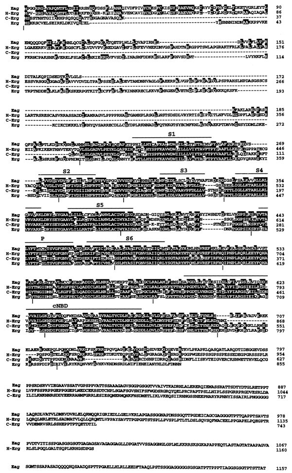
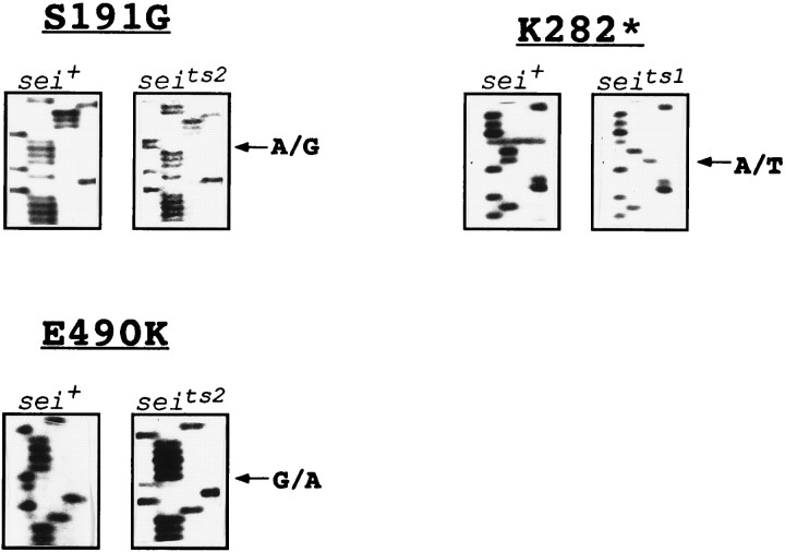
The eag family of K+ channels contains three known subtypes: eag, elk, and erg. Genes representing the first two subtypes have been identified in flies and mammals, whereas the third subtype has been defined only by the human HERG gene, which encodes an inwardly rectifying channel that is mutated in some cardiac arrhythmias. To establish the predicted existence of a Drosophila gene in the erg subfamily and to learn more about the structure and biological function of channels within this subfamily, we undertook a search for the Drosophila counterpart of HERG. Here we report the isolation and characterization of the Drosophila erg gene. We show that it corresponds with the previously identified seizure (sei) locus, mutations of which cause a temperature-sensitive paralytic phenotype associated with hyperactivity in the flight motor pathway. These results yield new insights into the structure and evolution of the eag family of channels, provide a molecular explanation for the sei mutant phenotype, and demonstrate the important physiological roles of erg-type channels from invertebrates to mammals.


Figures

Fig. 1.
Fig. 1.
Amino acid sequence of the Drosophila erg polypeptide and its alignment with other members of theeag family of K+ channel polypeptides. Identical residues are shaded in black. The approximate locations of the presumptive membrane-spanning regions (S1–S6), the pore region (P), and the region of homology to a cyclic nucleotide-binding domain (cNBD) are overlined. Gaps in the alignment are indicated by dashes. Vertical bars beneath the alignment mark the positions of introns in theDrosophila erg genomic sequence. The Drosophila eag sequence and the human HERG sequence have been published previously (Warmke et al., 1991; Warmke and Ganetzky, 1994). The nematode erg sequence (C-erg) was obtained by using the GCG Blast program to search the GenBank database (see Materials and Methods).
Fig. 2.
Fig. 2.
Northern blot analysis of poly(A+)-selected RNA isolated from wild-type (Canton-S) adults and hybridized with an erg cDNA probe.Left lane contains 4 μg of RNA; right lane, 6 μg. The position of size markers is indicated on theright.
Fig. 3.
Fig. 3.
Embryonic expression pattern of ergdetermined by tissue whole-mount in situ hybridization. Lateral view of a stage 16 embryo oriented with anterior to theleft and dorsal up. Expression of theerg transcript can be detected throughout the CNS, including the ventral nerve cord (vnc) and the brain hemispheres (br). Staining in salivary glands, as seen in this embryo, occurred only sporadically.
Fig. 4.
Fig. 4.
Cytological mapping of the erglocus by chromosomal in situ hybridization. The site of hybridization (arrow) relative to the cytological landmarks in numbered region 60 is shown. The hybridization signal lies directly on top of salivary bands 60B1–2.
Fig. 5.
Fig. 5.
Sequence analysis of mutational changes caused byseits1 andseits2 in theerg polypeptide. In all gels, the sequencing reactions are loaded (from the left) in the order G, A, T, C. Segments of sequence from the sense strand are shown. Two lesions are present in seits2, an A to G substitution at nucleotide position 571 of the ORF, resulting in a replacement of Ser by Gly at amino acid position 191, and a G to A substitution at nucleotide position 1468, resulting in a replacement of Glu by Lys at amino acid position 490. Inseits1, there is an A to T substitution at nucleotide position 844, resulting in the change of a Lys codon to a stop codon at amino acid position 282.

References

    1. Atkinson N, Robertson G, Ganetzky B. A structural component of calcium-activated potassium channels encoded by the Drosophila slo locus. Science. 1991;253:551–555. - PubMed
    1. Bruggeman A, Pardo LA, Stuhmer W, Pongs O. Ether a-go-go encodes a voltage-gated channel permeable to K+ and Ca2+ and modulated by cAMP. Nature. 1993;365:445–448. - PubMed
    1. Butler AS, Tsunoda S, McCobb DP, Wei A, Salkoff L. mSlo, a complex mouse gene encoding “maxi” calcium-activated potassium channels. Science. 1993;261:221–224. - PubMed
    1. Cavener DC, Ray SC. Eukaryotic start and stop translation sites. Nucleic Acids Res. 1991;19:3185–3192. - PMC - PubMed
    1. Curran ME, Splawski I, Timothy KW, Vincent M, Green ED, Keating MT. A molecular basis for cardiac arrhythmia: HERG mutations cause long QT syndrome. Cell. 1995;80:795–804. - PubMed

Publication types

Associated data

LinkOut - more resources