. 1997 Feb 1;17(3):1055-63.
doi: 10.1523/JNEUROSCI.17-03-01055.1997.

Role of NMDA and non-NMDA ionotropic glutamate receptors in traumatic spinal cord axonal injury

Affiliations

Role of NMDA and non-NMDA ionotropic glutamate receptors in traumatic spinal cord axonal injury

S K Agrawal et al. J Neurosci. .

Abstract

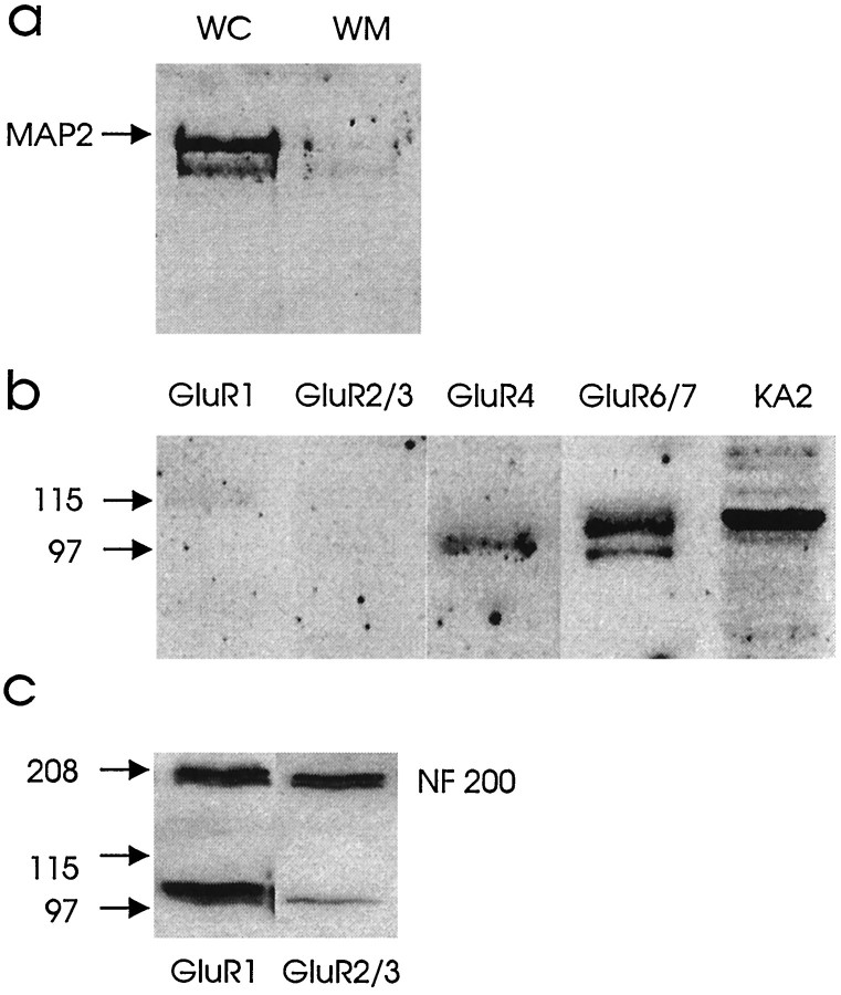
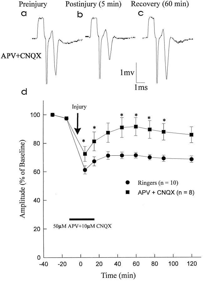
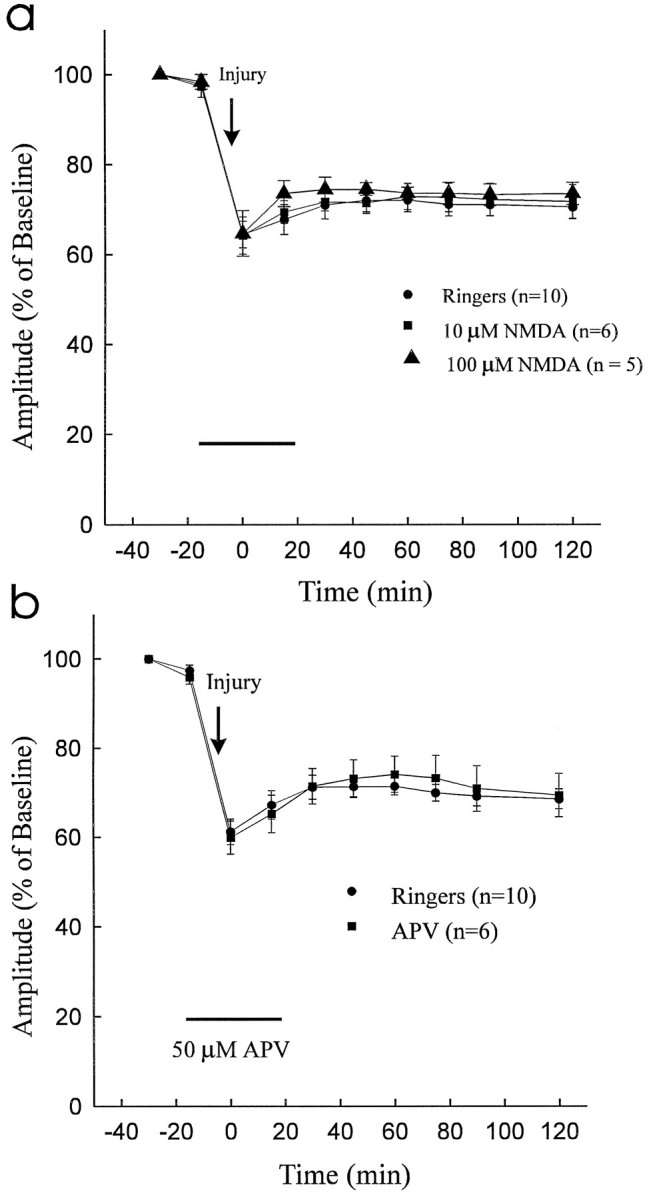
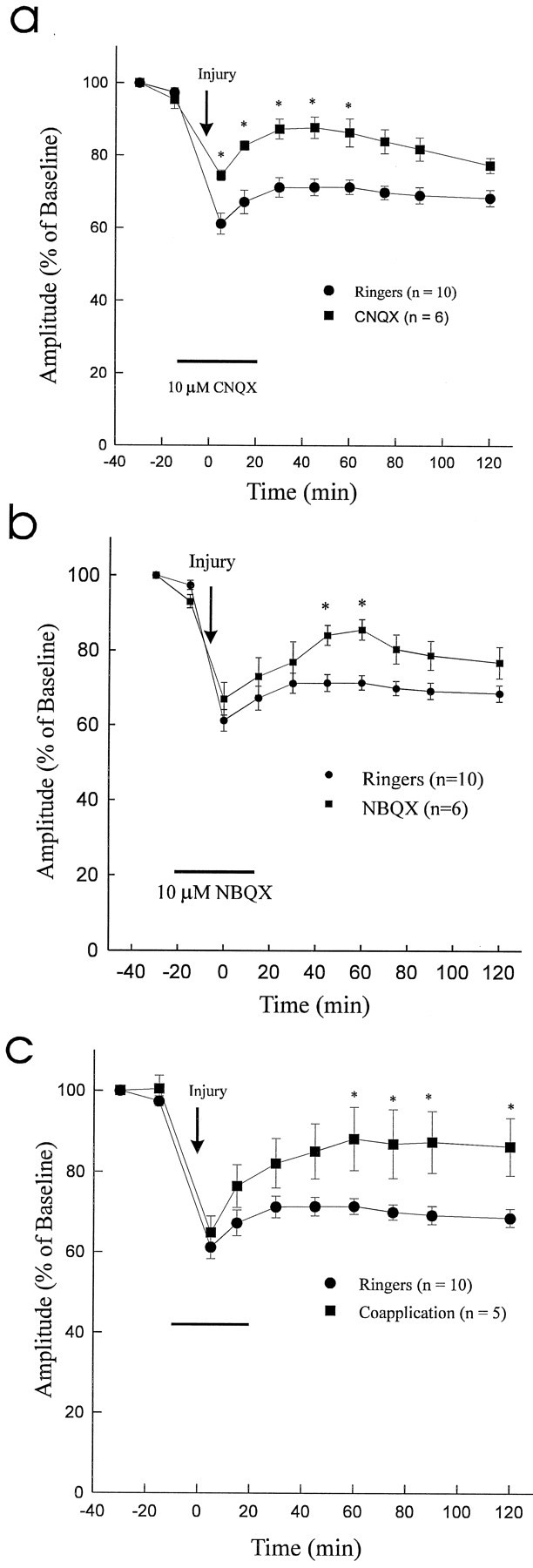
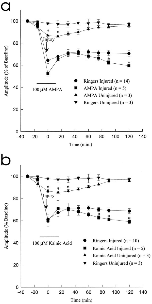
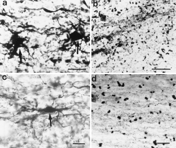
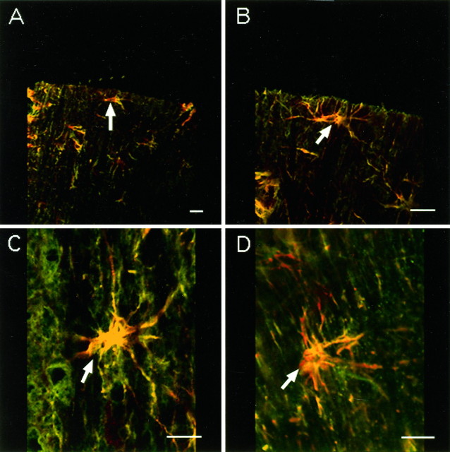
We examined the role of glutamatergic mechanisms in acute injury to rat spinal cord white matter. Compound action potentials (CAPs) were recorded from isolated dorsal column segments in vitro. Under control conditions (Ringer's solution), the CAPs decreased to 71.4 +/- 2.0% of preinjury values after compression injury with a clip exerting a closing force of 2 g. The combination of the NMDA receptor blocker APV (50 microM) and the AMPA/kainate (KA) receptor blocker CNQX (10 microM) resulted in significantly improved recovery of CAP amplitude postinjury; however, the NMDA receptor antagonist APV alone did not enhance postinjury recovery, and infusion of NMDA (10 microM) did not affect recovery of the CAPs. In contrast, the AMPA/KA receptor blockers NBQX (10 microM) or CNQX (10 microM) significantly enhanced the recovery of CAP amplitude postinjury. The agonists AMPA (100 microM) or KA (100 microM) resulted in significant attenuation of CAP amplitude postinjury. Coapplication of AMPA/KA plus NBQX and CNQX was also associated with improved functional recovery. After incubation with AMPA and KA, Co(2+)-positive glia were visualized in spinal cord white matter. Similar results were seen after compressive injury but not in control cords. Immunohistochemistry and Western blot analysis demonstrated AMPA (GluR4)- and KA (GluR6/7 and KA2)-positive astrocytes in spinal cord white matter. In summary, non-NMDA ionotropic glutamate receptors seem to be involved in the pathophysiology of traumatic spinal cord injury. The presence of AMPA (GluR4) and KA (GluR6/7 and KA2) receptors on periaxonal astrocytes suggests a role for these cells in glutamatergic white matter injury.


Figures

Fig. 6.
Fig. 6.
a, Western blot illustrating MAP2 presence in whole spinal cord (WC) and exclusion in an isolated dorsal column white matter preparation (WM). b, Spinal cord white matter (dorsal column) homogenates (10 μg protein) were subjected to SDS-PAGE and immunoblotted with antibodies to GluR1, GluR2/3, GluR4, GluR6/7, and KA2. As illustrated,GluR4, GluR6/7, and KA2 were detected in spinal cord white matter. c, These immunoblots show equal amounts of protein loading with NF 200 and positive controls for GluR1 and GluR2/3using olfactory bulb.
Fig. 1.
Fig. 1.
Effect of ionotropic glutamate receptor blockade on recovery of CAP amplitude after compressive injury. APV (50 μm; NMDA receptor blocker) and CNQX (10 μm; AMPA/KA receptor blocker) (APV+CNQX) were administered in combination and compared with control Ringer’s solution (Ringers). a–c, Representative CAP waveforms from the APV+CNQX group recorded preinjury, 5 min postinjury, and 60 min postinjury (during washout with Ringer’s solution). d, Graph of normalized CAP amplitude versus time. Combined administration of APV (50 μm) and CNQX (10 μm) resulted in improved CAP amplitude recovery postinjury (significant differences in CAP amplitude shown by asterisks). At 60 min postinjury, the CAP amplitude in the APV + CNQX group had recovered to 91.5 ± 6.4% of preinjury values as compared with theRingers group (71.4 ± 2.0% of preinjury).
Fig. 2.
Fig. 2.
a, Administration of NMDA (10 μm and 100 μm) did not attenuate CAP recovery after traumatic injury. b, The NMDA receptor antagonist APV (50 μm) did not improve recovery of CAP amplitude after compressive injury to the dorsal column segment.
Fig. 3.
Fig. 3.
Effect of 10 μmCNQX or NBQX or coapplication of AMPA/KA plus antagonists (CNQX and NBQX) on CAP recovery after 2 g clip compression injury.a, Graph of normalized CAP amplitude versus time (significant differences in CAP amplitude between two groups are depicted by asterisks). At 60 min postinjury, the recovery of CAP amplitude in the CNQX group (86.5 ± 3.9% of preinjury) significantly exceeded that of the control Ringer’s solution group (Ringers) (71.4 ± 2.0% of preinjury; p < 0.05). b, Graph of normalized CAP amplitude versus time (significant differences in CAP amplitude between two groups are depicted by asterisks). At 60 min postinjury the recovery of CAP amplitude in theNBQX group (85.6 ± 2.7% of preinjury) significantly exceeded that of the control Ringers group (71.4 ± 2.0% of preinjury; p < 0.05).c, Graph of normalized CAP amplitude versus time (significant differences in CAP amplitude between two groups are depicted by asterisks). At 60 min postinjury the recovery of CAP amplitude in the coapplication group (88.2 ± 7.8% of preinjury) significantly exceeded that of the controlRingers group (71.4 ± 2.0% of preinjury;p < 0.05).
Fig. 4.
Fig. 4.
Effect of AMPA (100 μm) and kainic acid (100 μm) on CAP amplitude after 2 gclip compression injury. Both AMPA and Kainic Acid resulted in significant exacerbation of traumatic axonal injury when compared with control Ringer’s solution group (Ringers) (asterisks denote significant differences at p < 0.05).
Fig. 5.
Fig. 5.
Longitudinal sections through spinal cord dorsal column stained for AMPA/KA receptors by the Co2+uptake technique. Scale bar, 20 μm. a, Two darkly stained astrocytes are shown (arrows). Section incubated with 100 μm kainic acid and 100 μm AMPA before histochemical processing. b, Control section (without AMPA/KA stimulation) of spinal cord dorsal column white matter showing background staining only. c, A darkly stained astrocyte is shown (arrow) in injured dorsal column (untreated with AMPA/KA). d, A dorsal column white matter preparation treated with CNQX and NBQX after clip compression injury showing no Co2+ uptake staining.
Fig. 7.
Fig. 7.
The presence of GluR4 (A, B), GluR6/7 (C), and KA2 (D) immunoreactivity (FITC labeling shown as green) in 10 μm sections of thoracic spinal cord dorsal column. Double labeling with GFAP (Texas Red) confirmed these immunopositive cells to be astrocytes (arrows). Scale bars, 50 μm.

References

    1. Agrawal SK, Fehlings MG. Mechanisms of calcium mediated axonal injury after spinal cord trauma. Soc Neurosci Abstr. 1995;21:381.
    1. Agrawal SK, Fehlings MG. Mechanisms of secondary injury to spinal cord axons in vitro: role of Na+, Na+–K+ ATPase, the Na+–H+ exchanger, and the Na+–Ca++ exchanger. J Neurosci. 1996;16:545–552. - PMC - PubMed
    1. Ascher P, Nowak L. Quisqualate- and kainate-activated channels in mouse central neurons in culture. J Physiol (Lond) 1988;399:227–245. - PMC - PubMed
    1. Barres BA. New roles for glia. J Neurosci. 1991;11:3685–3694. - PMC - PubMed
    1. Barres BA, Koroshetz WJ, Swartz KJ, Chun LLY, Corey DP. Ion channel expression by white matter glia: the O-2A glial progenator cell. Neuron. 1990;4:507–524. - PubMed

Publication types

MeSH terms

Substances