. 1997 Jan 6;185(1):55-64.
doi: 10.1084/jem.185.1.55.

Macrophage-dependent apoptosis of CD4+ T lymphocytes from HIV-infected individuals is mediated by FasL and tumor necrosis factor

Affiliations

Macrophage-dependent apoptosis of CD4+ T lymphocytes from HIV-infected individuals is mediated by FasL and tumor necrosis factor

A D Badley et al. J Exp Med. .

Abstract

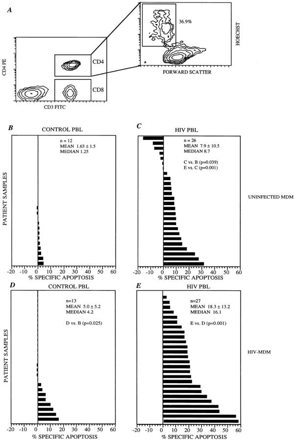
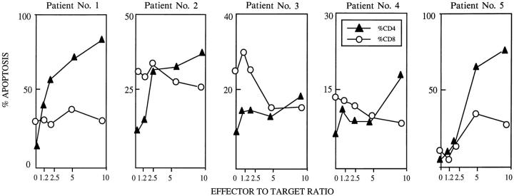
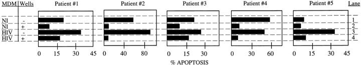
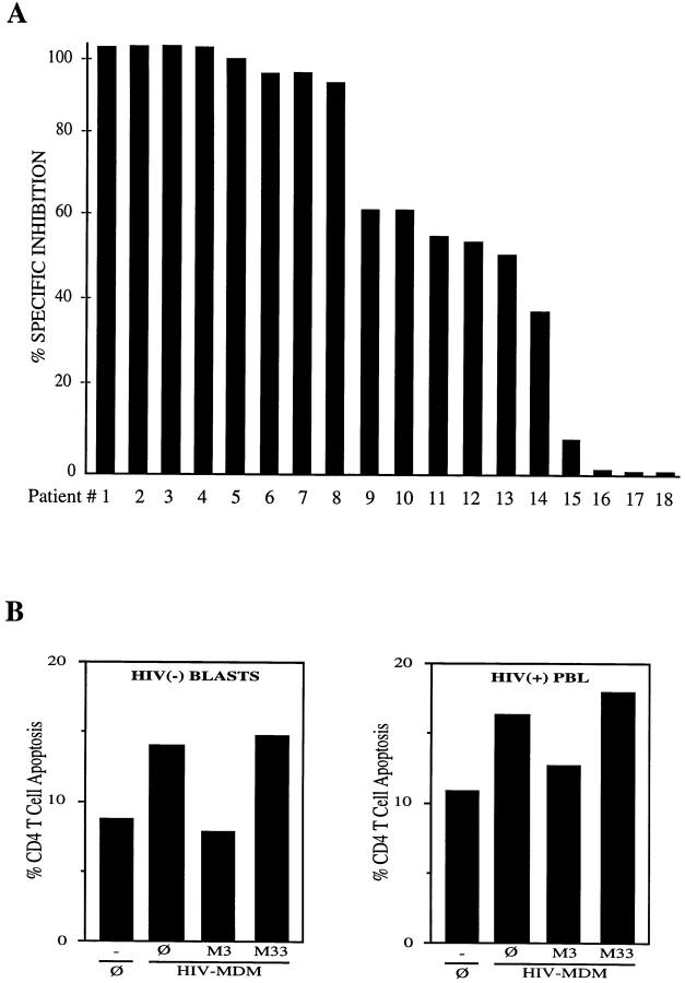
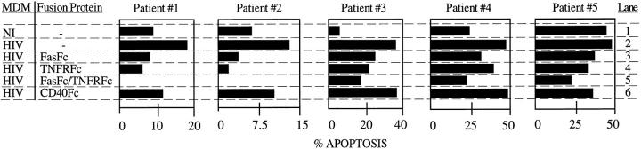
Apoptosis of bystander uninfected CD4+ T lymphocytes by neighboring HIV-infected cells is observed in cell culture and in lymphoid tissue of HIV-infected individuals. This study addresses whether antigen-presenting cells such as human macrophages mediate apoptosis of CD4+ T cells from HIV-infected individuals. Uninfected human macrophages, and to a larger degree, HIV-infected macrophages mediate apoptosis of T cells from HIV-infected, but not from uninfected control individuals. This macrophage-dependent killing targets CD4+, but not CD8+ T lymphocytes from HIV-infected individuals, and direct contact between macrophages and lymphocytes is required. Additional analyses indicated that the apoptosis-inducing ligands, FasL and tumor necrosis factor (TNF), mediate this macrophage-induced apoptosis of CD4+ T cells. These results support a role for macrophage-associated FasL and TNF in the selective depletion of CD4+ T cells in HIV-infected individuals.


Figures

Figure 1
Figure 1
Apoptosis of CD4+ T cells from HIV seropositive individuals by uninfected and HIV-infected macrophages. (A) Analysis of apoptosis can be performed separately on CD4+ T lymphocytes (CD3 FITC positive, CD4+ PE positive cells) and on CD8+ lymphocytes (CD3 FITC positive, CD4+ PE negative cells). In the plot depicted, 36.9% of cells have decreased forward angle light scatter and increased Hoechst specific fluorescence, indicating that these cells are apoptotic within the CD4 population (right). (B and C) Uninfected macrophages (UNINFECTED MDM) were used as effector cells at 5:1 effector/target ratio against PBL from either HIV seronegative (B) or infected individuals (C). Mean spontaneous apoptosis was 7.8 ± 4.9% (B), and 23.1 ± 15.1% (C). (D and E) The same experiments repeated with HIV-infected MDM (HIV-MDM) against HIV seronegative (D) or seropositive (E) individuals. Median spontaneous apoptosis was 8.1 ± 4.8% (D), and 23.0 ± 14.8 (E). In B–E, percent specific apoptosis was calculated by subtracting the amount of apoptosis from PBLs cultured in medium alone from the amount of apoptosis when the same PBLs were coincubated with macrophages as indicated.
Figure 2
Figure 2
HIV-infected macrophages induce selective apoptosis of CD4+ T cells from HIV-infected individuals. 106 PBL from HIV-infected and HIV seronegative healthy individuals were coincubated with varying amounts (0, 1, 2, 5, and 10 × 106/well) of HIV-infected macrophages for 36 h, and stained with anti-CD3 FITC, anti-CD4+ PE, and Hoechst 33342. The observed amounts of apoptosis for CD4 lymphocytes (CD3+, CD4+) and CD8 lymphocytes (CD3+, CD4) are shown. Spontaneous apoptosis is defined as the degree of apoptosis of PBL incubated in the absence of MDM.
Figure 3
Figure 3
Contact between HIV-MDM and PBL is required to induce apoptosis of T lymphocytes. Macrophages that were either HIV- or mock- infected were coincubated with PBL from HIV seropositive individuals with either the two cell populations in direct contact (−), or with contact interrupted (+) by a semipermeable transwell. The mean spontaneous apoptosis in these experiments was 11.0 ± 3.4%.
Figure 4
Figure 4
(A) MDM induced apoptosis of CD4+ T lymphocytes is partially mediated by FasL. PBL from HIV-infected individuals were incubated alone or with HIV-infected macrophages at an effector/target ratio of 5:1, either in the absence or presence of blocking (Fab′ M3) or nonblocking (Fab′ M33) anti-Fas antibodies (10 μg/well). In all cases, Fab′ M33 did not significantly alter the level of apoptosis seen in the PBL coincubated with macrophages without antibody. In order to determine the contribution of FasL to MDM killing of PBLs, percent specific inhibition was calculated as follows: ([apoptosis of PBL coincubated with MDM − apoptosis of PBL coincubated with Fab′3 and MDM] [apoptosis of PBL coincubated with MDM − apoptosis of PBL incubated alone]). The mean spontaneous apoptosis was 20.7 ± 13.7%. (B) Syngeneic apoptosis of susceptible CD4+ T cells by HIV-infected MDM. (Left) Lymphocyte blasts from an HIV seronegative individual (HIV(−) BLASTS) were incubated with 3 × 10⁶ syngeneic HIV-infected MDM (HIV-MDM) either alone () or in the presence of blocking (Fab′ M3) or nonblocking (Fab′ M33) antibodies (10 μg/well). Spontaneous apoptosis was calculated by incubating PBL blasts without macrophages (). Incubation with crosslinked agonistic M3 anti-Fas antibody lead to 27% CD4+ T cell apoptosis. Freshly isolated PBLs had a spontaneous level of CD4+ T cell apoptosis of 2% which was not modified (3%) upon coincubation with HIV-infected MDM. (Right) Freshly isolated PBLs from an HIV-infected individual (HIV(+) PBL) were incubated with syngeneic HIV-infected MDMs (3 × 10⁶) in the absence or presence of Fab M3 or M33 (10 μg/ml) antibodies. Cross-linked M3 antibodies induced 15% of CD4+ T cell apoptosis.
Figure 5
Figure 5
FasL and TNF participate in MDM-induced apoptosis of CD4+ T cells from HIV-infected individuals. 5 × 10⁶ uninfected (NI) or HIV- infected (HIV) macrophages were incubated with 106 PBL from five different HIV-infected individuals in the presence or absence of 20 μg FasFc, TNFRFc, or CD40Fc or 10 μg of each of TNFRFc and FasFc for 48 h. Analysis of CD4+ T cell apoptosis was done as described in Fig. 1 A. Observed amounts of apoptosis for CD4 T cells are shown. Mean spontaneous apoptosis was 23.5 ± 9.8%.

References

    1. Krammer PH, Behrmann I, Daniel P, Dhein J, Debatin K-M. Regulation of apoptosis in the immune system. Curr Opin Immunol. 1994;6:279–289. - PubMed
    1. Alderson MR, Tough TW, Braddy S, Davis T, Smith, Roux E, Schooley K, Miller RE, Lynch DH. Regulation of apoptosis and T cell activation by Fas-specific mAb. Int Immunol. 1994;6:1799–1806. - PubMed
    1. Dhein J, Walczak H, Bäumler C, Debatin K-M, Krammer PH. Autocrine T-cell suicide mediated by APO-1/(Fas/CD95) Nature (Lond) 1995;373:438–441. - PubMed
    1. Brunner T, Mogil RJ, LaFace D, Yoo NJ, Mahboubi A, Echeverri F, Martin SJ, Force WR, Lynch DH, Ware CF, Green DR. Cell-autonomous Fas (CD95)/Fasligand interaction mediates activation-induced apoptosis in T-cell hybridomas. Nature (Lond) 1995;373:441–444. - PubMed
    1. Alderson MR, Tough TW, Davis-Smith T, Braddy S, Falk B, Schooley KA, Goodwin RG, Smith CA, Ramsdell F, Lynch DH. Fas ligand mediates activationinduced cell death in human T lymphocytes. J Exp Med. 1995;181:71–77. - PMC - PubMed

MeSH terms