. 1997 Jan 6;185(1):71-9.
doi: 10.1084/jem.185.1.71.

Scleroderma autoantigens are uniquely fragmented by metal-catalyzed oxidation reactions: implications for pathogenesis

Affiliations

Scleroderma autoantigens are uniquely fragmented by metal-catalyzed oxidation reactions: implications for pathogenesis

L Casciola-Rosen et al. J Exp Med. .

Abstract

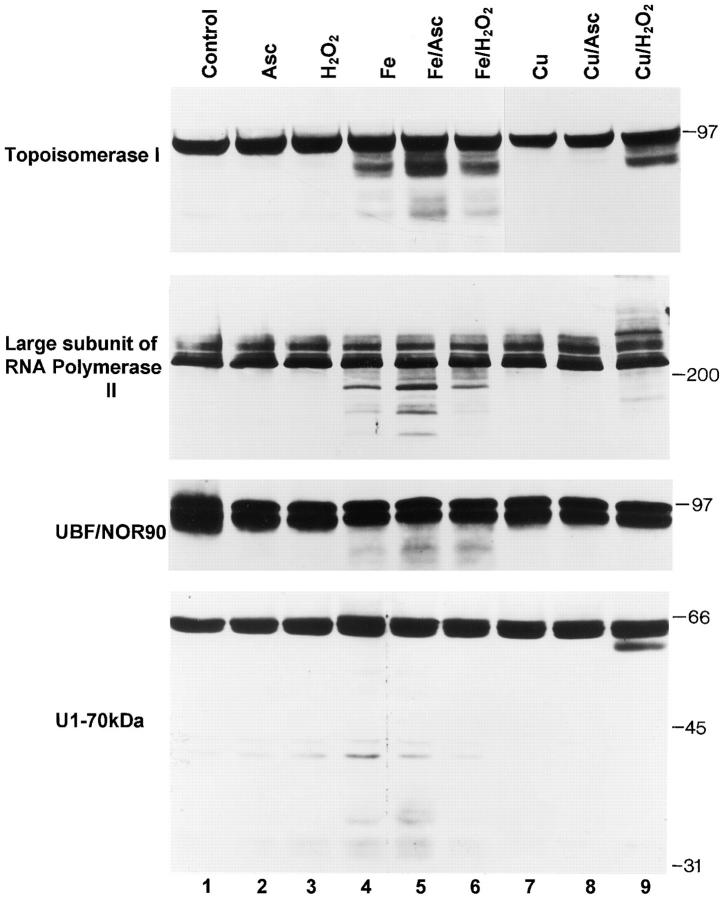
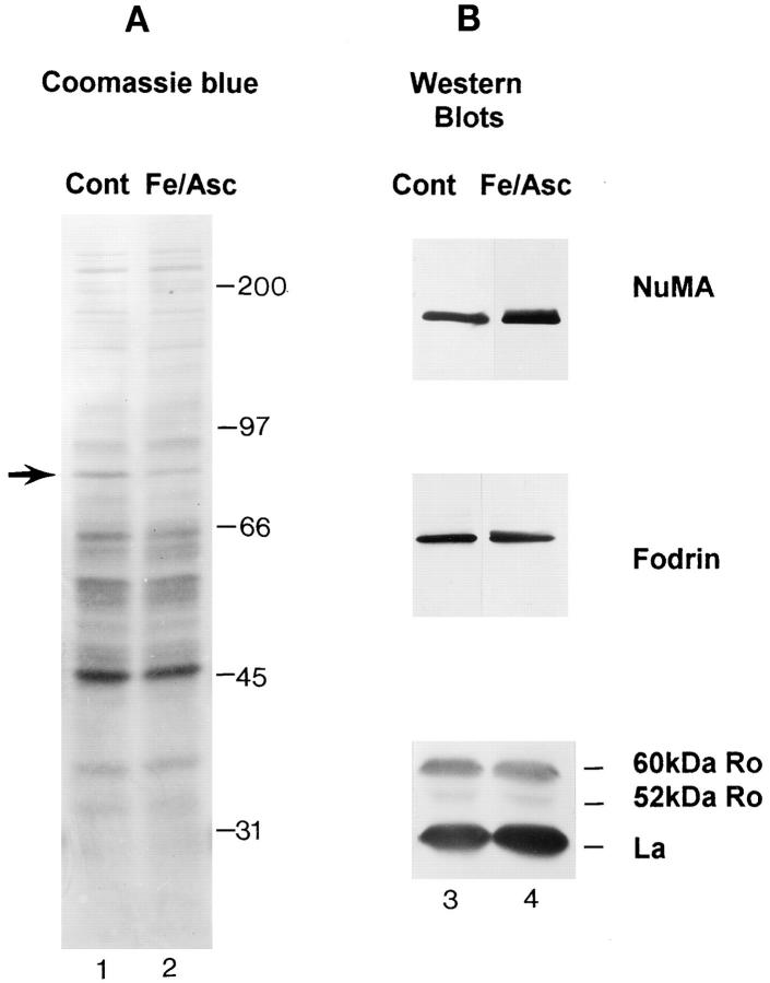
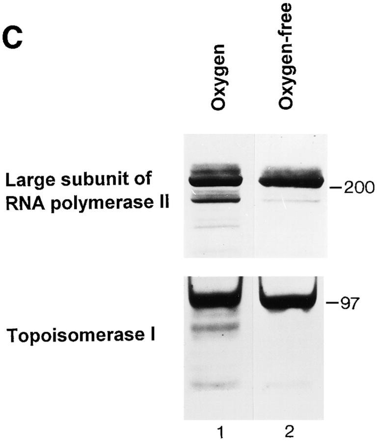
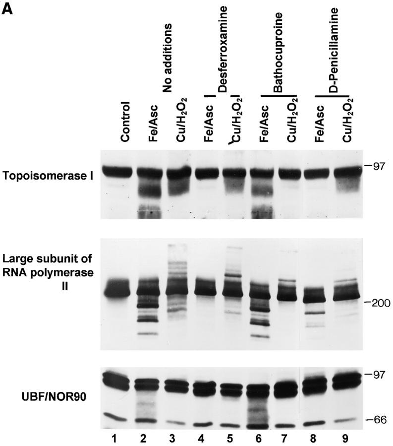
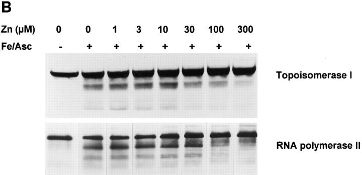
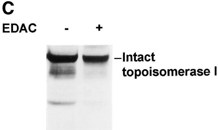
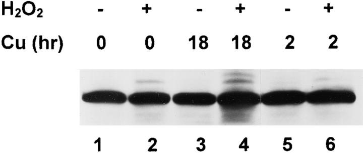
The observation that revelation of immunocryptic epitopes in self antigens may initiate the autoimmune response has prompted the search for processes which induce novel fragmentation of autoantigens as potential initiators of autoimmunity. The reversible ischemia reperfusion which characterizes scleroderma has focused attention on reactive oxygen species as molecules which might induce autoantigen fragmentation. We demonstrate that several of the autoantigens targeted in diffuse scleroderma are uniquely susceptible to cleavage by reactive oxygen species, in a metal-dependent manner. Multiple features of the fragmentation reaction and its inhibition indicate that these autoantigens possess metal-binding sites, which focus metal-catalyzed oxidation reactions (and consequent fragmentation) to specific regions of the antigens. These data suggest that the autoantibody response in scleroderma is the immune marker of unique protein fragmentation, induced by ischemia reperfusion in the presence of appropriate metals, and focus attention on abnormal metal status as a potential pathogenic principle in this disease.


Figures

Figure 1
Figure 1
Several scleroderma autoantigens are uniquely fragmented by Fe/ascorbate or Cu/H2O2 oxidation reactions. HeLa lysates were prepared as described in the Materials and Methods section, and metal-catalyzed oxidation reactions were performed by adding the following: no additions (lane 1), 1.7 mM ascorbate (lane 2), 1 mM H2O2 (lane 3), 100 μM Fe(II)SO4 (lane 4), 100 μM Fe(II)SO4 + 1.7 mM ascorbate (lane 5), 100 μM Fe(II)SO4 + 1 mM H2O2 (lane 6), 100 μM Cu(II)SO4 (lane 7), 100 μM Cu(II)SO4 + 1.7 mM ascorbate (lane 8) and 100μM Cu(II)SO4 + 1 mM H2O2 (lane 8). Samples were immunoblotted with the sera denoted on the left side of each panel. Equal amounts of protein were electrophoresed in the lanes of each panel. Migration positions of molecular weight standards are indicated on the right.
Figure 2
Figure 2
Fragmentation of scleroderma autoantigens by oxidation is a highly specific event. (A and B) Equal protein amounts of control HeLa lysates (lanes 1 and 3), and HeLa lysates incubated with 100 μM Fe + 1.7 mM ascorbate (lanes 2 and 4) were electrophoresed. (A) Gels were stained with Coomassie blue to visualize the total protein profile of each sample. Oxidation catalyzed by Fe/ascorbate caused no striking alterations in the Coomassie pattern, with the exception of the decreased staining of an ∼85 kD protein (arrow). Migration positions of the molecular weight standards are indicated to the right of lane 2. (B) Electrophoresed proteins were immunoblotted with patient sera recognizing NuMA, La, and 52 and 60-kD Ro, or a monoclonal antibody to fodrin (Chemicon International Inc., Temecula, CA). None of these antigens were fragmented by Fe/ascorbate oxidation reactions (lanes 3 and 4). (C) Fragmentation of RNA polymerase II and topoisomerase I is decreased in O2-depleted lysates. O2 depletion was performed as described in Materials and Methods, before adding 100 μM Fe and 1.7 mM ascorbate. Equal protein amounts were electrophoresed and immunoblotted with antibodies to RNA polymerase II and topoisomerase I.
Figure 2
Figure 2
Fragmentation of scleroderma autoantigens by oxidation is a highly specific event. (A and B) Equal protein amounts of control HeLa lysates (lanes 1 and 3), and HeLa lysates incubated with 100 μM Fe + 1.7 mM ascorbate (lanes 2 and 4) were electrophoresed. (A) Gels were stained with Coomassie blue to visualize the total protein profile of each sample. Oxidation catalyzed by Fe/ascorbate caused no striking alterations in the Coomassie pattern, with the exception of the decreased staining of an ∼85 kD protein (arrow). Migration positions of the molecular weight standards are indicated to the right of lane 2. (B) Electrophoresed proteins were immunoblotted with patient sera recognizing NuMA, La, and 52 and 60-kD Ro, or a monoclonal antibody to fodrin (Chemicon International Inc., Temecula, CA). None of these antigens were fragmented by Fe/ascorbate oxidation reactions (lanes 3 and 4). (C) Fragmentation of RNA polymerase II and topoisomerase I is decreased in O2-depleted lysates. O2 depletion was performed as described in Materials and Methods, before adding 100 μM Fe and 1.7 mM ascorbate. Equal protein amounts were electrophoresed and immunoblotted with antibodies to RNA polymerase II and topoisomerase I.
Figure 3
Figure 3
Metal chelators, zinc, or EDAC strongly inhibit or abolish fragmentation of the scleroderma autoantigens induced by both Fe/ascorbate and Cu/H2O2. The following additions were made to equal protein amounts of HeLa cell lysate, before immunoblotting with the indicated antibodies: (A) none (lane 1), 100 μM Fe + 1.7 mM ascorbate (lanes 2, 4, 6, and 8); 100 μM Cu + 1 mM H2O2 (lanes 3, 5, 7, and 9); 1 mM desferroxamine (lanes 4 and 5); 200 μM bathocuproine disulfonate (lanes 6 and 7), or 1 mM D-penicillamine (lanes 8 and 9). The band migrating at 66 kD in the UBF/NOR90 immunoblot is nonspecific. (B) Increasing concentrations of ZnCl2 (0–300 μM) were added to HeLa lysates before addition of 100 μM Fe + 1.7 mM ascorbate. The IC50 value for inhibition of Fe/ascorbate-induced fragmentation of topoisomerase I or RNA polymerase II large subunit was 30–50 μM. (C) HeLa lysates were incubated in the absence (−) or presence (+) of 5 mM EDAC, before adding 100 μM Fe and 1.7 mM ascorbate. (A–C) Equal protein amounts were loaded in each lane. Results are representative of two to six separate experiments.
Figure 3
Figure 3
Metal chelators, zinc, or EDAC strongly inhibit or abolish fragmentation of the scleroderma autoantigens induced by both Fe/ascorbate and Cu/H2O2. The following additions were made to equal protein amounts of HeLa cell lysate, before immunoblotting with the indicated antibodies: (A) none (lane 1), 100 μM Fe + 1.7 mM ascorbate (lanes 2, 4, 6, and 8); 100 μM Cu + 1 mM H2O2 (lanes 3, 5, 7, and 9); 1 mM desferroxamine (lanes 4 and 5); 200 μM bathocuproine disulfonate (lanes 6 and 7), or 1 mM D-penicillamine (lanes 8 and 9). The band migrating at 66 kD in the UBF/NOR90 immunoblot is nonspecific. (B) Increasing concentrations of ZnCl2 (0–300 μM) were added to HeLa lysates before addition of 100 μM Fe + 1.7 mM ascorbate. The IC50 value for inhibition of Fe/ascorbate-induced fragmentation of topoisomerase I or RNA polymerase II large subunit was 30–50 μM. (C) HeLa lysates were incubated in the absence (−) or presence (+) of 5 mM EDAC, before adding 100 μM Fe and 1.7 mM ascorbate. (A–C) Equal protein amounts were loaded in each lane. Results are representative of two to six separate experiments.
Figure 4
Figure 4
Topoisomerase I is fragmented in intact keratinocytes chronically exposed to 20 μM Cu. Confluent monolayers of human foreskin keratinocytes were cultured for 18 h (lanes 3 and 4) or 2 h (lanes 5 and 6) in keratinocyte growth medium supplemented with 20 μM CuSO4. Control cultures were maintained in the absence of added Cu (lanes 1 and 2). Before harvesting the cells, 2 mM H2O2 was added to some of the cultures (lanes 2, 4, and 6), but not others (lanes 1 3, and 5), for 30 min. Cells were subsequently harvested and immunoblotted with anti-topoisomerase I serum as described in Fig. 1. Equal amounts of protein were electrophoresed in each lane. Results are representative of those obtained in three separate experiments.
Figure 5
Figure 5
Metal ions and topoisomerase I are concentrated in punctate intranucleolar structures. (A) HeLa cells were fixed in 1% lead acetate, and subsequently stained with 1% ammonium sulfide. Bright field microscopy demonstrates intense staining of punctate intranucleolar structures (nucleolini). (B) HeLa cells were stained with a monospecific human serum recognizing topoisomerase I and FITC-goat anti– human IgG, and were examined by confocal fluoresence microscopy. Diffuse nuclear staining, as well as punctate intranucleolar structures (nucleolini), are seen. Similar results were obtained with six other monospecific topoisomerase I sera. Nucleolini were never stained when similar experiments were performed using sera obtained from healthy individuals, or from Ro/La-positive lupus patients. Bar, 10 μM.

Similar articles

Cited by

References

    1. Radic MZ, Weigert M. Genetic and structural evidence for antigen selection of anti-DNA antibodies. Annu Rev Immunol. 1994;12:487–520. - PubMed
    1. Diamond B, Katz JB, Paul E, Aranow C, Lustgarten D, Scharff MD. The role of somatic mutation in the pathogenic anti-DNA response. Annu Rev Immunol. 1992;10:731–757. - PubMed
    1. Sercarz EE, Lehmann PV, Ametani A, Benichou G, Miller A, Moudgil K. Dominance and crypticity of T cell antigenic determinants. Annu Rev Immunol. 1993;11:729–766. - PubMed
    1. Lanzavecchia A. How can cryptic epitopes trigger autoimmunity? . J Exp Med. 1995;181:1945–1948. - PMC - PubMed
    1. Gammon G, Sercarz EE. How some T cells escape tolerance induction. Nature (Lond) 1989;342:183–185. - PubMed

Publication types