

Developmental regulation of VDJ recombination by the core fragment of the T cell receptor alpha enhancer
- PMID: 8996249
- PMCID: PMC2196107
- DOI: 10.1084/jem.185.1.131
Developmental regulation of VDJ recombination by the core fragment of the T cell receptor alpha enhancer
Abstract
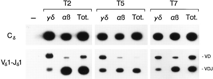
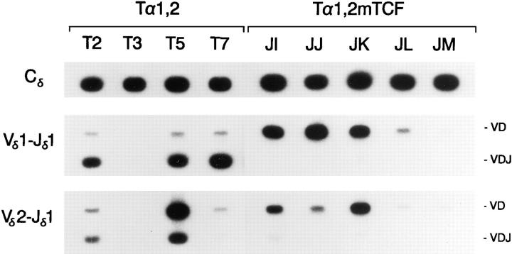
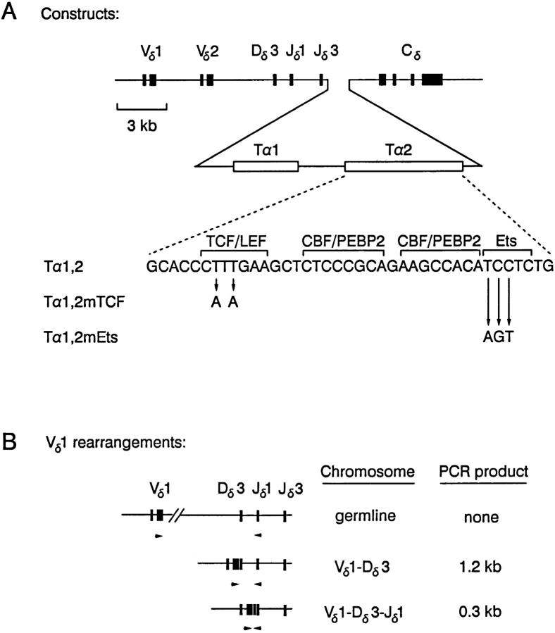
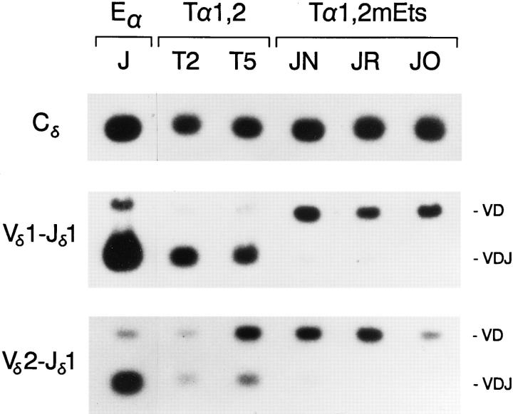
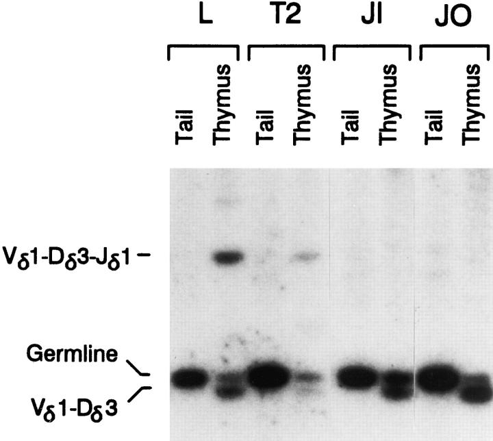
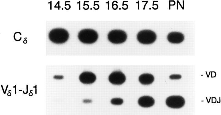
The role of T cell receptor alpha enhancer (E alpha) cis-acting elements in the developmental regulation of VDJ recombination at the TCR alpha/delta locus was examined in transgenic mice containing variants of a minilocus VDJ recombination substrate. We demonstrate that the 116-bp T alpha 1,2 core enhancer fragment of the 1.4-kb E alpha is sufficient to activate the enhancer-dependent step of minilocus rearrangement, and that within T alpha 1,2, intact binding sites for TCF/LEF and Ets family transcription factors are essential. Although minilocus rearrangement under the control of the 1.4-kb E alpha initiates at fetal day 16.5 and is strictly limited to alpha beta T cells, we find that rearrangement under the control of T alpha 1,2 initiates slightly earlier during ontogeny and occurs in both gamma delta and alpha beta T cells. We conclude that the core fragment of E alpha can establish accessibility to the recombinase in developing thymocytes in vivo in a fashion that is dependent on the binding of TCF/LEF and Ets family transcription factors, but that these and other factors that bind to the E alpha core cannot account for the precise developmental onset of accessibility that is provided by the intact E alpha. Rather, our data suggests a critical role for factors that bind E alpha outside of the core T alpha 1,2 region in establishing the precise developmental onset of TCR alpha rearrangement in vivo.
Figures
Similar articles
-
Regulation of T cell receptor delta gene rearrangement by CBF/PEBP2.J Exp Med. 1997 Apr 7;185(7):1193-201. doi: 10.1084/jem.185.7.1193. J Exp Med. 1997. PMID: 9104806 Free PMC article.
-
Temporal and lineage-specific control of T cell receptor alpha/delta gene rearrangement by T cell receptor alpha and delta enhancers.J Exp Med. 1994 Jun 1;179(6):1913-21. doi: 10.1084/jem.179.6.1913. J Exp Med. 1994. PMID: 8195717 Free PMC article.
-
A delta T-cell receptor deleting element transgenic reporter construct is rearranged in alpha beta but not gamma delta T-cell lineages.Mol Cell Biol. 1995 Dec;15(12):7022-31. doi: 10.1128/MCB.15.12.7022. Mol Cell Biol. 1995. PMID: 8524269 Free PMC article.
-
Accessibility control of T cell receptor gene rearrangement in developing thymocytes. The TCR alpha/delta locus.Immunol Res. 2000;22(2-3):127-35. doi: 10.1385/IR:22:2-3:127. Immunol Res. 2000. PMID: 11339350 Review.
-
Enforcing order within a complex locus: current perspectives on the control of V(D)J recombination at the murine T-cell receptor alpha/delta locus.Immunol Rev. 2004 Aug;200:224-32. doi: 10.1111/j.0105-2896.2004.00155.x. Immunol Rev. 2004. PMID: 15242408 Review.
Cited by
-
T cell receptor (TCR) alpha/delta locus enhancer identity and position are critical for the assembly of TCR delta and alpha variable region genes.Proc Natl Acad Sci U S A. 2003 Mar 4;100(5):2598-603. doi: 10.1073/pnas.0437943100. Epub 2003 Feb 25. Proc Natl Acad Sci U S A. 2003. PMID: 12604775 Free PMC article.
-
Cryptic signals and the fidelity of V(D)J joining.Mol Cell Biol. 1997 Jun;17(6):3125-36. doi: 10.1128/MCB.17.6.3125. Mol Cell Biol. 1997. PMID: 9154811 Free PMC article.
-
Positive and negative regulation of V(D)J recombination by the E2A proteins.J Exp Med. 1999 Jan 18;189(2):289-300. doi: 10.1084/jem.189.2.289. J Exp Med. 1999. PMID: 9892611 Free PMC article.
-
TCRα rearrangements identify a subgroup of NKL-deregulated adult T-ALLs associated with favorable outcome.Leukemia. 2018 Jan;32(1):61-71. doi: 10.1038/leu.2017.176. Epub 2017 Jun 8. Leukemia. 2018. PMID: 28592888 Clinical Trial.
-
Genomic analysis offers insights into the evolution of the bovine TRA/TRD locus.BMC Genomics. 2014 Nov 19;15(1):994. doi: 10.1186/1471-2164-15-994. BMC Genomics. 2014. PMID: 25408163 Free PMC article.
References
-
- Fowlkes BJ, Pardoll DM. Molecular and cellular events of T cell development. Adv Immunol. 1989;44:207–264. - PubMed
-
- Rothenberg EV. The development of functionally responsive T cells. Adv Immunol. 1992;51:85–214. - PubMed
-
- Kisielow P, von Boehmer H. Development and selection of T cells: facts and puzzles. Adv Immunol. 1995;58:87–209. - PubMed
-
- Wu L, Scollay R, Egerton M, Pearse M, Spangrude GJ, Shortman K. CD4 expressed on earliest T-lineage precursor cells in the adult murine thymus. Nature (Lond) 1991;349:71–74. - PubMed
Publication types
MeSH terms
Substances
Grants and funding
LinkOut - more resources
Full Text Sources
Molecular Biology Databases
