. 1997 Jan 6;185(1):153-63.
doi: 10.1084/jem.185.1.153.

Loss of ATP diphosphohydrolase activity with endothelial cell activation

Affiliations

Loss of ATP diphosphohydrolase activity with endothelial cell activation

S C Robson et al. J Exp Med. .

Abstract

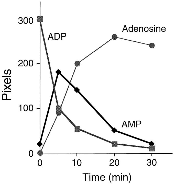
Quiescent endothelial cells (EC) regulate blood flow and prevent intravascular thrombosis. This latter effect is mediated in a number of ways, including expression by EC of thrombomodulin and heparan sulfate, both of which are lost from the EC surface as part of the activation response to proinflammatory cytokines. Loss of these anticoagulant molecules potentiates the procoagulant properties of the injured vasculature. An additional thromboregulatory factor, ATP diphosphohydrolase (ATPDase; designated as EC 3.6.1.5) is also expressed by quiescent EC, and has the capacity to degrade the extracellular inflammatory mediators ATP and ADP to AMP, thereby inhibiting platelet activation and modulating vascular thrombosis. We describe here that the antithrombotic effects of the ATPDase, like heparan sulfate and thrombomodulin, are lost after EC activation, both in vitro and in vivo. Because platelet activation and aggregation are important components of the hemostatic changes that accompany inflammatory diseases, we suggest that the loss of vascular ATPDase may be crucial for the progression of vascular injury.


Figures

Figure 1
Figure 1
Effect of pEC on platelet aggregation induced by ADP and ADP-β-S (an ADP analog resistant to ATPDase activity). Platelets underwent comparable aggregation responses after stimulation with 1 μM ADP and 5 μM ADP-β-S. Addition of pEC abrogated platelet responses to ADP alone, but had minimal effects on ADP-β-S stimulated platelet aggregation. These data indicate a functional ATPDase associated with EC is responsible, at least in part, for the inhibitory effects on platelet aggregation in vitro.
Figure 2
Figure 2
Hydrolysis of [14C]ADP to AMP by EC-associated ATPDase. Radiolabeled ADP hydrolysis to AMP, and consequent catalysis to adenosine by pEC, was measured by TLC of supernatants from EC cultures. ADP was rapidly degraded and the radio-label appeared as AMP initially, and then adenosine over a time period of 30 min.
Figure 3
Figure 3
ATPDase antiaggregatory activity is modulated by EC responses to TNFα in vitro. Activation of quiescent porcine EC by 10 ng/ml human recombinant TNFα from 1 to 8 h in vitro resulted in rapid loss of the activated EC antiaggregatory phenotype at the time of testing. This was noted by the development of a permissive environment for platelet activation in response to 5 μM ADP in vitro. After TNFα stimulation of EC, reconstitution of functional antiplatelet aggregatory properties was observed by 18 h.
Figure 4
Figure 4
ATPDase enzymatic activity after EC activation by TNFα. EC ATPDase activity was determined by measuring inorganic phosphate release from ADP and ATP. An inhibitory effect was maximal by 4 h after stimulation of pEC by TNFα as depicted here for ADP (data in graph are expressed as means and SD; normality test passed). Statistical analysis confirmed significant differences to control quiescent EC values at both 2 and 4 h after EC activation; *P <0.005, Mann Whitney Rank Sum Test. Experiments studying ATPDase activity by [14C]ADP hydrolysis gave similar results (data not shown).
Figure 5
Figure 5
Effect of EC activation upon ATPDase antigen expression. (a) Western blotting. The polyclonal antibody recognized ATPDase in human EC preparations purified from quiescent- and TNFαstimulated cells by Western blotting. We did not observe significant diminution of ATPDase antigen expression, nor evidence for proteolytic degradation, despite the documented reduction in enzyme activity at this time point. (b) Immunocytofluorometric analysis. These plots demonstrate high levels of surface expression of CD39 on quiescent HUVEC (bold line, anti-CD39; faint line, isotype control mAb) (A). The expression of CD39 epitopes on the EC surface was largely unaltered after TNFα stimulation (10 ng/ml; B) or direct oxidative stress with H2O2 (100 μM; C). Cells were analyzed by flow cytometry as described in Materials and Methods.
Figure 5
Figure 5
Effect of EC activation upon ATPDase antigen expression. (a) Western blotting. The polyclonal antibody recognized ATPDase in human EC preparations purified from quiescent- and TNFαstimulated cells by Western blotting. We did not observe significant diminution of ATPDase antigen expression, nor evidence for proteolytic degradation, despite the documented reduction in enzyme activity at this time point. (b) Immunocytofluorometric analysis. These plots demonstrate high levels of surface expression of CD39 on quiescent HUVEC (bold line, anti-CD39; faint line, isotype control mAb) (A). The expression of CD39 epitopes on the EC surface was largely unaltered after TNFα stimulation (10 ng/ml; B) or direct oxidative stress with H2O2 (100 μM; C). Cells were analyzed by flow cytometry as described in Materials and Methods.
Figure 6
Figure 6
Effects of oxidative stress on EC ATPDase activity. (a) ATPDase biochemical activity assay. The decreased capacity of pEC directly perturbed by oxidative stress to express ATPDase activity was demonstrated by estimation of phosphate release from supplemental ADP by the malachite green technique. Exogenous xanthine oxidase (XO, 100 mU/ml) and xanthine (X, 100 μM) markedly inhibited pEC ATPDase activity in a statistically significant manner after 2 h oxidant exposure (*P = 0.002; Mann Whitney Rank Sum Test). This effect could be abrogated by the supplemental antioxidants superoxide dismutase (SOD; 330 U/ml) and catalase (1,000 U/ml), confirming the purely oxidant nature of the inhibition (data are expressed as means and standard deviations; normality test passed for graphical representation). (b) Platelet inhibitory properties of ATPDase. Further evidence for the loss of ATPDase functional activity after exposure to oxidants was derived from the demonstration that pEC exposed to such reactions for 2 h were unable to inhibit platelet aggregation responses to ADP 5 μM.
Figure 6
Figure 6
Effects of oxidative stress on EC ATPDase activity. (a) ATPDase biochemical activity assay. The decreased capacity of pEC directly perturbed by oxidative stress to express ATPDase activity was demonstrated by estimation of phosphate release from supplemental ADP by the malachite green technique. Exogenous xanthine oxidase (XO, 100 mU/ml) and xanthine (X, 100 μM) markedly inhibited pEC ATPDase activity in a statistically significant manner after 2 h oxidant exposure (*P = 0.002; Mann Whitney Rank Sum Test). This effect could be abrogated by the supplemental antioxidants superoxide dismutase (SOD; 330 U/ml) and catalase (1,000 U/ml), confirming the purely oxidant nature of the inhibition (data are expressed as means and standard deviations; normality test passed for graphical representation). (b) Platelet inhibitory properties of ATPDase. Further evidence for the loss of ATPDase functional activity after exposure to oxidants was derived from the demonstration that pEC exposed to such reactions for 2 h were unable to inhibit platelet aggregation responses to ADP 5 μM.
Figure 7
Figure 7
Maintenance of pEC-associated ATPDase activity by antioxidants after TNFα stimulation. Superoxide dismutase (SOD; Cu-Zn form, 330 U/ml); the hydrogen peroxide scavenger catalase (1,000 U/ml) and the 21-aminosteroid des-methyl tirilazad (U74389; final concentration 5 μM) were again used as antioxidants. Such interventions could protect against the statistically significant TNFα-mediated inhibitory changes in pEC ATPDase activity, and had minimal positive effects on quiescent pEC ATPDase levels. Inhibition of ATPDase activity following TNFα activation was consistently abrogated by these antioxidants (data are expressed as mean and standard deviations).
Figure 8
Figure 8
Loss of ATPDase activity during reperfusion injury in vivo. Upper panels show the extent of enzyme histochemical activity (cerium chloride method) within representative glomeruli of rat kidneys which were (a) freshly harvested, or (b) subjected to 1 h of ischemia and a further hour of reperfusion. Compared to the moderate to dense enzyme expression by rat vascular EC in control glomeruli (a), reperfusion injury and associated oxidative stress reduced ATPDase activity to negligible levels (b); arrows indicate neutrophils within capillary loops of kidney subjected to reperfusion injury. Lower panels are corresponding Hematoxylin- and Eosin-stained sections, showing (c) good preservation of glomerular structure and absence of leukocytes, and (d) association of reperfusion injury with focal platelet microthrombi which fill some capillary loops and the presence of neutrophils (arrows). All panels ×200.

References

    1. Preissner KT. Anticoagulant potential of endothelial cell membrane components. Haemostasis. 1988;18:271–306. - PubMed
    1. Ihrcke NS, Wrenshall LE, Lindman BJ, Platt JL. Role of heparan sulfate in immune system-blood vessel interactions. Immunol Today. 1993;14:500–505. - PubMed
    1. Esmon CT. Protein-S and protein-C–biochemistry, physiology, and clinical manifestation of deficiencies. Trends Cardiovasc Med. 1992;2:214–219. - PubMed
    1. Nawroth PP, Handley DA, Esmon CT, Stern DM. Interleukin 1 induces endothelial cell procoagulant while suppressing cell-surface anticoagulant activity. Proc Natl Acad Sci USA. 1986;83:3460–3464. - PMC - PubMed
    1. Saadi S, Holzknecht RA, Patte CP, Stern DM, Platt JL. Complement mediated regulation of tissue factor activity in endothelium. J Exp Med. 1995;182:1807–1814. - PMC - PubMed

Publication types

MeSH terms

Associated data