. 1997 Jan;109(1):93-104.
doi: 10.1085/jgp.109.1.93.

Subunit stoichiometry of human muscle chloride channels

Affiliations

Subunit stoichiometry of human muscle chloride channels

C Fahlke et al. J Gen Physiol. 1997 Jan.

Abstract

Voltage-gated Cl- channels belonging to the ClC family appear to function as homomultimers, but the number of subunits needed to form a functional channel is controversial. To determine subunit stoichiometry, we constructed dimeric human skeletal muscle Cl- channels in which one subunit was tagged by a mutation (D136G) that causes profound changes in voltage-dependent gating. Sucrose-density gradient centrifugation experiments indicate that both monomeric and dimeric hClC-1 channels in their native configurations exhibit similar sedimentation properties consistent with a multimeric complex having a molecular mass of a dimer. Expression of the heterodimeric channel in a mammalian cell line results in a homogenous population of Cl- channels exhibiting novel gating properties that are best explained by the formation of heteromultimeric channels with an even number of subunits. Heteromultimeric channels were not evident in cells cotransfected with homodimeric WT-WT and D136G-D136G constructs excluding the possibility that functional hClC-1 channels are assembled from more than two subunits. These results demonstrate that the functional hClC-1 unit consists of two subunits.


Figures

Figure 6
Figure 6
Cotransfection of WT–WT and D136G–D136G dimer constructs. (A, C, and E) Current recordings in cells cotransfected with equal quantities of WT–WT and D136G–D136G DNAs elicited with voltage steps to between −165 and +75 mV in 80 mV-steps from a holding potential of 0 mV. Each voltage step is followed by a fixed test pulse to either −105 mV (A and C) or −135 mV (E). (B, D, and F) Simulated current records of superimposed WT and D136G Cl currents in ratios of 5:1 (B), 2:1 (D), or 1:2 (F ).
Figure 7
Figure 7
Activation curves for homomultimeric and heteromultimeric channels. (A, C, and E) Current responses to voltage steps between −165 mV and + 75 mV in 40-mV steps followed by a fixed test pulse to −105 mV. Measurements were performed on cells expressing either WT (A), WT–D136G (C), or D136G (E) channels. (B, D, and F ) Voltage dependence of the normalized current amplitude measured either at the beginning (filled circles, Ipeak) or at the end of (open squares, Iss) the −105-mV test pulse for WT (D), WT- D136G (E), and D136G (F ) channels. Each point represents mean ± SD (n = 4).
Figure 1
Figure 1
Expression of WT–WT and D136G –D136G homodimeric constructs. Responses to voltage steps from a holding potential of 0 mV to between −165 mV and +85 mV in 50-mV steps. Test pulses are followed by a voltage step to −125 mV. (A–D) Current recordings made from cells expressing either WT hClC-1 (A), the WT–WT dimeric construct (B), D136G hClC-1 (C), or the D136G –D136G dimeric construct (D).
Figure 3
Figure 3
Properties of WT–D136G heteromultimeric hClC-1 channels. (A) Current recordings from a cell transiently transfected with the WT–D136G dimeric construct elicited with voltage steps to between −165 and +85 mV in 50-mV steps. Each voltage step is followed by a −125-mV test pulse. (B) Current recordings from a cell transiently transfected with the D136G–WT dimeric construct elicited with voltage steps to between −165 and +85 mV in 50-mV steps. Each voltage step is followed by a −125-mV test pulse. (C) Normalized current recordings from WT, WT–D136G, or D136G hClC-1 expressing cells at a test potential of −165 mV. The dotted lines represent the addition of WT and D136G current traces scaled such that the peak current amplitude and the amplitude at the end of the test step are identical to the normalized WT–D136G recording. (D) Normalized current recordings from WT, WT–D136G, or D136G hClC-1 expressing cells at a test potential of −115 mV. The dotted lines represent the addition of WT and D136G current traces scaled such that the peak current amplitude and the amplitude at the end of the test step are identical to the normalized WT–D136G recording.
Figure 8
Figure 8
Quantitative analysis of WT:D136G homodimer and monomer co-expression. (A) Current recordings from a cell cotransfected with WT–WT and D136G–D136G homodimer constructs. Responses to voltage steps between −165 and +115 mV in 40-mV steps are illustrated. Each voltage step is followed by a fixed −105-mV test pulse. (B) Voltage dependence of the instantaneous current amplitude (filled circles, Ipeak) and the current amplitude at the end of the −105-mV test pulse (open squares, Iss) in WT–WT:D136G– D136G cotransfected cells. (C) Current amplitudes shown in (Fig. 7 C) corrected for the contribution of Cl currents conducted by D136G homomultimeric channels as described in the text. (D) Current recordings from a cell cotransfected with WT and D136G monomer constructs. Responses to voltage steps between −165 and +15 mV in 60-mV steps are illustrated. Each voltage step is followed by a fixed −105-mV test pulse. (E) Voltage dependence of the instantaneous current amplitude (filled circles, Ip eak) and the current amplitude at the end of the −105-mV test pulse (open squares, Iss) in WT:D136G co-transfected cells. (F ) Current amplitudes corrected for the contribution of Cl currents conducted by D136G homomultimeric channels as described in the text.
Figure 2
Figure 2
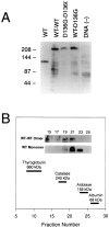
Biochemical characterization of hClC-1 dimers. (A) Western blot of recombinant hClC-1 constructs and sham transfected cells [DNA (–)]. The migration of molecular mass (in kD) standards are shown on the left of the panel. (B) Sucrose density gradient centrifugation experiment using WT and WT–WT hClC-1 constructs. The location of size standards are indicated by thick horizontal lines positioned above the corresponding fractions from the sucrose gradient. Inset: autoradiographs showing immunodetection of hClC-1 proteins in various gradient fractions (WT– WT dimer; WT monomer). Maximal immunoreactivity was seen in fractions 19–21 for WT–WT dimer, and 21–23 for WT monomer.
Figure 4
Figure 4
Homogenous gating properties of WT–D136G. Activation was examined in current recordings (dotted lines) elicited by voltage steps from −100 mV to between −65 and −25 mV in 20-mV steps for WT–hClC-1 (A), WT–D136G (B), and D136G (C). Solid lines represent fits with I(t) = a 1 e −t/τ1 + a 2 e −t/τ2 + c in A and I(t) = a 1 e −t/τ1 + c in B where a 1 and a 2 are amplitude terms and τ1 and τ2 are time constants. Currents shown for D136G are not fitted.
Figure 5
Figure 5
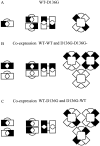
Possible configurations of heterodimeric and homodimeric hClC-1 channels. WT hClC-1 is illustrated by a black square and D136G by a white square. Each subunit has a single pore forming region indicated by the concave face. (A) Assembly of WT–D136G heterodimers into heterotetramers with either one or two pores is shown. (B) Assembly of tetrameric channels by the combination of WT–WT and D136G–D136G homodimers is shown. If hClC-1 is a tetramer, then homodimeric constructs would be expected to form a mixture of homotetrameric and heterotetrameric channels. (C) Assembly of WT–D136G and D136G – WT heterotandems into various tetrameric arrangements.
Figure 9
Figure 9
Co-expression of heterotandem hClC-1 constructs in Xenopus oocytes. (A) Recordings from oocytes expressing WT– D136G. Currents were elicited by voltage steps from −165 to +35 mV in 40-mV steps from a holding potential of −30 mV. (B) Recordings from oocytes expressing D136G–WT. Currents were elicited by voltage steps from −165 to +35 mV in 40-mV steps from a holding potential of −30 mV. (C) Recordings made from oocytes expressing both WT–D136G and D136G–WT. (D) Plot of the fraction of steady-current (Iss/Ipeak) vs voltage obtained from four to five cells. Lines without data points represent averaged data plots for D136G (solid line, n = 4) and WT (dotted line, n = 4) determined under identical conditions in oocytes.

References

    1. Adachi S, Uchida S, Ito H, Hata M, Hiroe M, Marumo F, Sasaki S. Two isoforms of a chloride channel predominantly expressed in thick ascending limb of Henle's loop and collecting ducts of rat kidney. J Biol Chem. 1994;269:17677–17683. - PubMed
    1. Bauer CK, Steinmeyer K, Schwarz JR, Jentsch TJ. Completely functional double-barreled chloride channel expressed from a single TorpedocDNA. Proc Natl Acad Sci USA. 1991;88:11052–11056. - PMC - PubMed
    1. Chahine M, George AL, Jr, Zhou M, Ji S, Sun W, Barchi RL, Horn R. Sodium channel mutations in paramyotonia congenita uncouple inactivation from activation. Neuron. 1994;12:281–294. - PubMed
    1. Deal KK, Lovinger DM, Tamkun MM. The brain Kv1.1 potassium channel: in vitro and in vivo studies on subunit assembly and posttranslational processing. J Neurosci. 1994;14:1666–1676. - PMC - PubMed
    1. Fahlke C, Rosenbohm A, Mitrovic N, George AL, Jr, Rüdel R. Mechanism of voltage-dependent gating in skeletal muscle chloride channels. Biophys J. 1996;71:695–706. - PMC - PubMed

Publication types

Substances