. 1997 Feb 15;17(4):1197-205.
doi: 10.1523/JNEUROSCI.17-04-01197.1997.

Cloning, characterization, and expression of a G-protein-coupled receptor from Lymnaea stagnalis and identification of a leucokinin-like peptide, PSFHSWSamide, as its endogenous ligand

Affiliations

Cloning, characterization, and expression of a G-protein-coupled receptor from Lymnaea stagnalis and identification of a leucokinin-like peptide, PSFHSWSamide, as its endogenous ligand

K J Cox et al. J Neurosci. .

Abstract

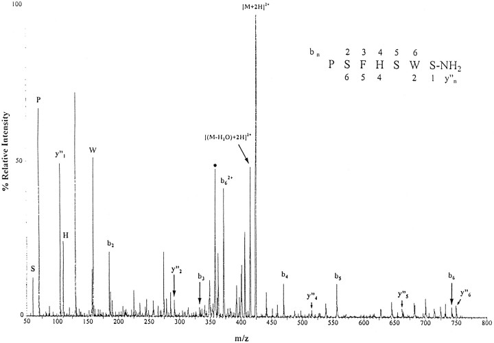
Neuropeptides are known to be important signaling molecules in several neural systems of the pond snail Lymnaea stagnalis. Although the functions of these peptides have been studied in many neurons, the nature of the postsynaptic signal transduction is mainly unknown. The cloning and characterization of neuropeptide receptors in Lymnaea thus would be very valuable in further elucidating peptidergic pathways. Indirect evidence suggests that these neuropeptides operate via G-protein-coupled mechanisms indicating the presence of G-protein-coupled receptors as the initial postsynaptic targets. Here we describe the cloning of a neuropeptide receptor from Lymnaea and the isolation of an endogenous ligand. This peptide, PSFHSWSamide, belongs to the leucokinin family of peptides, and, thus, this Lymnaea receptor is the first example of a leucokinin-like neuropeptide receptor, representing a new subfamily of G-protein-coupled neuropeptide receptors.


Figures

Fig. 1.
Fig. 1.
Nucleotide and deduced amino acid sequence ofLymnaea GRL104 receptor cDNA cloned in pBluescript BS. A methionine at position 374 indicates the start of the open reading frame of 1287 bp, which translates into 429 amino acids. Putative transmembrane regions are underlined and labeled I–VII. Cysteines suspected of being involved in a cysteine bridge between extracellular loops 2 and 3 are indicated byfilled circles. Arrowheads indicate possible N-linked glycosylation sites. The nucleotide sequence for GRL104 has been deposited into GenBank, accession number U84499.
Fig. 2.
Fig. 2.
Shown are transmembrane (TM) region amino acid alignments between GRL104 and other G-protein-coupled receptors: SK, human substance K receptor (Gerard et al., 1990); SP, rat substance P receptor (Yokota et al., 1989); NPY, human NPY Y1 receptor (Herzog et al., 1992); Musc, human m1 ACh receptor (Peralta et al., 1987); DA, human D1 dopamine receptor (Dearry et al., 1990); 5HT, human 5HT1d receptor (Hamblin and Metcalf, 1991). Shaded boxesindicate residues that are conserved among both classical and peptide receptors. Nonshaded boxes indicate residues that are conserved only among peptide receptors.
Fig. 3.
Fig. 3.
PCR analysis of CHO cell lines stably transfected with pcD104, using primers specific to pcD104. Lanes 1–6 contain PCR products from CHO cell lines KC2, KC4, KC5, KC6, KC7, and KC9. Lane 7 is a negative control using untransfected CHO-K1 cells as template. Lane 8 is a control PCR reaction with no template. Lane M contains size markers generated from λ bacteriophage cut byHindIII.
Fig. 4.
Fig. 4.
Purification of the endogenous ligand for GRL104. All assays were performed on a CHO cell line that had been stably transfected with pcD104 cDNA (CHO cell line GRL104 KC6). Thetop of each panel, A–C, gives the chromatogram for the HPLC purification indicated.A1, HPGPC fractionation of an extract of 500 Lymnaea brains;B1, rpHPLC fractionation of combined fractions 23 and 24 from A;C1, rpHPLC fractionation of fraction 29 from B. D, rpHPLC fractionation of combined fractions 36 and 37 from C. The bottom of each panel, A–C, shows the effect of the indicated HPLC fractions on intracellular calcium levels in the CHO cell line GRL104 KC6, using 2, 4, and 8 CNS equivalents forA2,B2,B3,C2, andC3, respectively.x-Axis numbers indicate HPLC fraction number. The calcium increases were calculated as increases in concentration above basal levels. On each HPLC fractionation,A2 shows the two fractions (of 33) that were active (mean of two determinations),B3 shows the fraction (of 54) that was active (mean of two determinations), andC3 shows the two fractions (of 45) that were active (one determination only to conserve enough of the fraction for the next stage of HPLC, seen in D).
Fig. 5.
Fig. 5.
Tandem mass spectrometric analysis of lymnokinin. Averaged daughter ion spectra of the purified lymnokinin, generated from the doubly charged parent ion species (M + 2H)2+ ofm/z 424 Da. The Roepstorff nomenclature is used to identify fragment ions (Roepstorff and Fohlman, 1984). The y” ions are formed by charge retention on C-terminal fragments, and the a and b ions are formed on N-terminal fragments. The measured protonated mass of the peptide [846.8 Da, as detected in stage 1 (ms 1) of the tandem MS analysis; data not shown] as well as the y”, a, and b ion series are in perfect agreement with the calculated protonated masses of the peptide PSFHSWSamide (846.9 Da as detected in ms 1; data not shown) and the corresponding y”, a, and b ion series. Theasterisk represents a62+,x-axis; m/z is mass to charge ratio.
Fig. 6.
Fig. 6.
Dose–response curve of the increase in intracellular calcium in GRL104 KC6 CHO cells elicited by synthetic lymnokinin. The calcium increases were calculated as increases in concentration above basal calcium levels. Each data point is the mean of three separate determinations. Error bars are ± SD.

References

    1. Benjamin PR, Burke JF. Alternative mRNA splicing of the FMRFamide gene and its role in neuropeptidergic signalling in a defined network. BioEssays. 1994;16:335–342. - PubMed
    1. Blackburn MB, Wagner RM, Shasanowitz J, Kochansky JP, Hunt DF, Raina AK. The isolation and identification of 3 diuretic kinins from the abdominal ventral nerve cord of adult Helicoverpa zea. J Insect Physiol. 1996;41:723–730.
    1. Blitz DM, Christie AE, Marder EM, Nusbaum MP. Distribution and effects of tachykinin-like peptides in the stomatogastric nervous system of the crab, Cancer borealis. J Comp Neurol. 1995;354:282–294. - PubMed
    1. Bloem LJ, Yu L. A time-saving method for screening cDNA or genomic libraries. Nucleic Acids Res. 1990;18:2830. - PMC - PubMed
    1. Chen Y, Veenstra JA, Davis NT, Hagedorn HH. A comparative study of leucokinin-immunoreactive neurons in insects. Cell Tissue Res. 1994;276:69–83. - PubMed

Publication types

Substances

Associated data