. 1997 Feb 15;17(4):1217-25.
doi: 10.1523/JNEUROSCI.17-04-01217.1997.

Characterization of a CNS cell line, CAD, in which morphological differentiation is initiated by serum deprivation

Affiliations

Characterization of a CNS cell line, CAD, in which morphological differentiation is initiated by serum deprivation

Y Qi et al. J Neurosci. .

Abstract

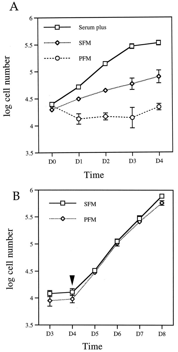
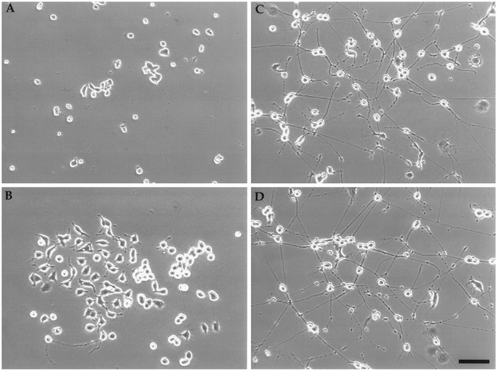
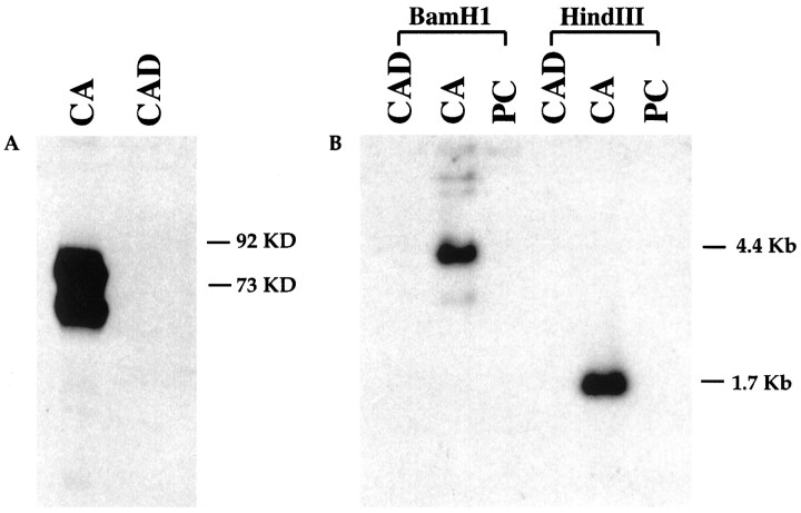
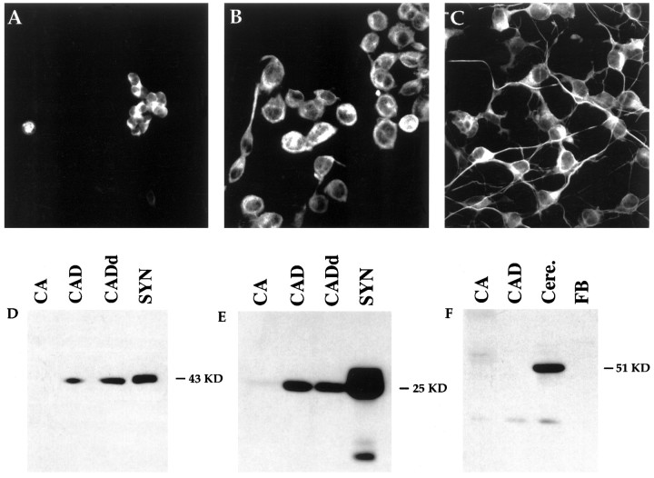
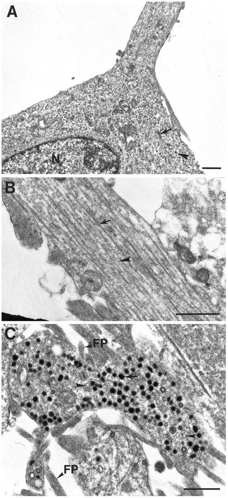
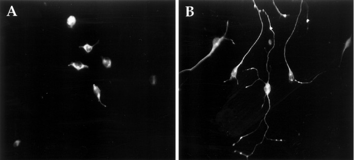
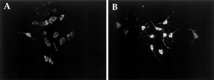
A CNS catecholaminergic cell line, Cath.a, was established by targeted oncogenesis in transgenic mice. Cath.a cells express neuronal properties but lack neuronal morphology. Here, we describe a variant of Cath.a, called CAD (Cath.a-differentiated), in which reversible morphological differentiation can be initiated by removal of serum or exogenously added protein from the medium. In serum- or protein-free media, CAD cells stop proliferating and extend long processes. Differentiated CAD cells can be maintained without serum or protein for at least 6 weeks. CAD cells are distinct from Cath.a cells; most significant, the original immortalizing oncogene, SV40 T antigen, was spontaneously lost. By immunostaining or immunoblotting, we show that CAD cells express neuron-specific proteins, such as class III beta-tubulin, GAP-43, SNAP-25, and synaptotagmin, but not GFAP. Ultrastructurally, processes from differentiated CAD cells have abundant parallel microtubules and intermediate filaments, and bear varicosities that contain both large dense-core vesicles/granules (120-160 nm) and smaller clear vesicles (60-80 nm). Additionally, CAD cells express enzymatically active tyrosine hydroxylase and accumulate L-DOPA. CAD cells exhibit biochemical and morphological characteristics of primary neurons and provide an unique tool for studying neuronal differentiation.


Figures

Fig. 2.
Fig. 2.
Reversible reduction of cell proliferation rate in SFM or PFM. A, Growth curve of CAD cells. Cells were grown in serum-containing medium; on day 0 (D0), they were changed into SFM or PFM or maintained in serum-containing medium for an additional 4 d. Values are the average of the cell number from four separate experiments. The cell number was obtained as described in Materials and Methods. B, Differentiated CAD cells resumed proliferation after serum addition. CAD cells were maintained in SFM or PFM for 4 d, after which FBS was added to a final concentration of 8% to the culture medium.Arrowhead indicates serum addition. Error bars indicate SEM (n = 4). Note: some of the error bars are too small to be visible.
Fig. 1.
Fig. 1.
Morphological differentiation can be induced in CAD cells. Phase-contrast photomicrograph of live cells: Cath.a cells in serum-containing medium (A); CAD cells in serum-containing medium (B), SFM (C), or PFM (D). In C and D, the cells were grown for 5 d in SFM or PFM. Note the beaded varicosities on the processes. Scale bar, 100 μm.
Fig. 3.
Fig. 3.
SV40 T antigen (Tag) is lost in CAD cells.A, Western blot analysis with Tag-specific polyclonal antibody. Each lane contains 50 μg of protein. Tag was detected in Cath.a cells (CA) as a doublet. No Tag was detected in CAD cells. B, Southern blot analysis with a32P-labeled Tag probe. Tag is only detected in Cath.a cells, but not in CAD or PC12 cells. PC, PC12 cells;KD, kilodalton.
Fig. 4.
Fig. 4.
CAD cells express neuron-specific proteins.A–C, Immunocytochemical staining with TuJ1 mAb against class III β-tubulin. Cath.a (A), undifferentiated CAD (B), and differentiated CAD (C) cells were all stained with TuJ1 antibody. D–F, Western blot with antibodies against GAP-43 (D), SNAP-25 (E), and GFAP (F). Fifty micrograms of Cath.a (CA), CAD, and differentiated CAD (CADd) cell protein were loaded in each lane. GAP-43 was detected in both CAD cells and rat brain synaptosome (SYN) extracts. GAP-43 protein was increased twofold after differentiation (CADd). However, no GAP-43 was detected in Cath.a cells. SNAP-25 was detected in both CAD cells and synaptosome extracts, but not in Cath.a cells (E). In F, Cath.a, CAD, and fibroblasts (FB) did not express the glial-specific protein GFAP, although extracts from mouse cerebellum (Cere.) had abundant GFAP.
Fig. 5.
Fig. 5.
Electron micrographs of differentiated CAD cells.A, Cell body and initial segment of a process in differentiated CAD cells. The cytoplasm contains ribosomes (arrowhead), mitochondria (asterisk), rough endoplasmic reticulum (arrow), and microtubules.B, A typical process is filled with parallel microtubules (arrow) and intermediate filaments (arrowhead). C, Dense-core vesicles (arrow) and clear vesicles (arrowhead) in terminal. FP, Filopodia; N, nucleus. Scale bars, 1 μm.
Fig. 6.
Fig. 6.
Immunocytochemical staining of CAD cells with synaptotagmin mAb. A, CAD cells grown in serum-containing medium. B, CAD cells grown for 4 d in PFM.
Fig. 7.
Fig. 7.
Expression of TH in CAD cells. Immunocytochemistry with monoclonal TH antibody showing cytoplasmic TH staining in both growing CAD (A) and differentiated CAD cells (B).

References

    1. Amano T, Richelson E, Nirenberg M. Neurotransmitter synthesis by neuroblastoma clones. Proc Natl Acad Sci USA. 1972;69:258–263. - PMC - PubMed
    1. Augusti-Tocco G, Sato G. Establishment of functional clonal lines of neurons from mouse neuroblastoma. Proc Natl Acad Sci USA. 1969;64:311–315. - PMC - PubMed
    1. Bogenmann E, Torres M, Matsushima H. constitutive N-myc gene expression inhibits trkA mediated neuronal differentiation. Oncogene. 1995;10:1915–1925. - PubMed
    1. Bottenstein JE, Sato GH. Growth of a rat neuroblastoma cell line in serum-free supplemented medium. Proc Natl Acad Sci USA. 1979;76:514–517. - PMC - PubMed
    1. Boulter CA, Wagner EF. The effects of v-src expression on the differentiation of embryonal carcinoma cells. Oncogene. 1988;2:207–214. - PubMed

Publication types

Substances