. 1997 Feb 15;17(4):1226-42.
doi: 10.1523/JNEUROSCI.17-04-01226.1997.

Occurrence and biosynthesis of endogenous cannabinoid precursor, N-arachidonoyl phosphatidylethanolamine, in rat brain

Affiliations

Occurrence and biosynthesis of endogenous cannabinoid precursor, N-arachidonoyl phosphatidylethanolamine, in rat brain

H Cadas et al. J Neurosci. .

Abstract

It has been suggested that anandamide (N-arachidonoylethanolamine), an endogenous cannabinoid substance, may be produced through Ca2+-stimulated hydrolysis of the phosphatidylethanolamine (PE) derivative N-arachidonoyl PE. The presence of N-arachidonoyl PE in adult brain tissue and the enzyme pathways that underlie its biosynthesis are, however, still undetermined. We report here that rat brain tissue contains both anandamide (11 +/- 7 pmol/gm wet tissue) and N-arachidonoyl PE (22 +/- 16 pmol/gm), as assessed by gas chromatography/mass spectrometry. We describe a N-acyltransferase activity in brain that catalyzes the biosynthesis of N-arachidonoyl PE by transferring an arachidonate group from the sn-1 carbon of phospholipids to the amino group of PE. We also show that sn-1 arachidonoyl phospholipids are present in brain, where they constitute approximately 0.5% of total phospholipids. N-acyltransferase activity is Ca2+ dependent and is enriched in brain and testis. Within the brain, N-acyltransferase activity is highest in brainstem; intermediate in cortex, striatum, hippocampus, medulla, and cerebellum; and lowest in thalamus, hypothalamus, and olfactory bulb. Pharmacological inhibition of N-acyltransferase activity in primary cultures of cortical neurons prevents Ca2+-stimulated N-arachidonoyl PE biosynthesis. Our results demonstrate, therefore, that rat brain tissue contains the complement of enzymatic activity and lipid substrates necessary for the biosynthesis of the anandamide precursor N-arachidonoyl PE. They also suggest that biosynthesis of N-arachidonoyl PE and formation of anandamide are tightly coupled processes, which may concomitantly be stimulated by elevations in intracellular Ca2+ occurring during neural activity.


Figures
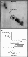
Fig. 1.
Fig. 1.
A, Identification of brainN-acyl PEs by bidimensional HPTLC. Phosphomolybdic acid staining revealed the presence of a lipid component, indicated by thearrow, with the chromatographic properties ofN-acyl PEs. Results are from one experiment, repeated once with identical results. B, Analytical approach used to confirm the identification of brain N-acyl PEs and to determine their molecular composition. Partially purifiedN-acyl PEs were digested with S. chromofuscus PLD to release the corresponding NAEs. These were subsequently purified by HPLC and analyzed by GC/MS.
Fig. 2.
Fig. 2.
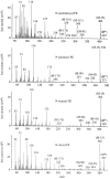
Electron-impact mass spectra of the TMS derivatives of synthetic anandamide (A),N-palmitoylethanolamine (B),N-stearoylethanolamine (C), andN-oleoylethanolamine (D).
Fig. 3.
Fig. 3.
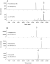
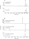
Identification ofN-arachidonoyl PE and other N-acyl PEs in brain by GC/MS. N-acyl PEs were digested withS. chromofuscus PLD, and the resulting NAEs were analyzed by GC/MS in the SIM mode as TMS derivatives. A, Anandamide, derived from the hydrolysis ofN-arachidonoyl PE; B, NAEs derived from the hydrolysis of N-palmitoyl PE,N-stearoyl PE, and N-oleoyl PE. Thearrows indicate the retention times of authentic standards. Results are from one experiment, representative of six.
Fig. 4.
Fig. 4.
Identification of anandamide and other NAEs in brain by GC/MS. The NAEs were purified chromatographically and analyzed by GC/MS in the SIM mode as TMS derivatives. A, Anandamide; B, additional NAEs. Thearrows indicate the retention times of authentic standards. Results are from one experiment, representative of six.
Fig. 5.
Fig. 5.
Brain particulate fractions contain an enzymatic activity that catalyzes the biosynthesis of N-acyl PEs. Particulate fractions were solubilized with the nonionic detergent, NP-40, and incubated either with 3 mm Ca2+(A) or without Ca2+ (B). The lipids were analyzed by HPTLC and visualized with phosphomolybdic acid. A lipid component with the chromatographic properties of syntheticN-acyl PEs, indicated by the arrow, was present only in the Ca2+-containing incubations. Results are from one experiment, representative of four.
Fig. 6.
Fig. 6.
Identification ofN-arachidonoyl PE and other N-acyl PEs produced by brain detergent-solubilized particulate fractions. The fractions (4.5 mg of protein) were incubated for 1 hr at 37°C, and the lipids were extracted. N-acyl PEs were partially purified and digested with PLD, and the NAEs produced in the digestions were analyzed by GC/MS as TMS derivatives. Complete mass spectra were obtained for each product. Results are from one experiment, repeated once with identical results.
Fig. 7.
Fig. 7.
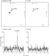
A Ca2+-dependentN-acyltransferase activity is present in particulate fractions. The detergent-solubilized fractions were incubated with 3 mm Ca2+ (A, C) or without Ca2+ (B, D), in a medium containing 1,2-di[14-C]palmitoyl PC as substrate. Radioactive products were identified by HPTLC (A, B) or by reversed-phase HPLC (C, D).
Fig. 8.
Fig. 8.
Identification of brainN-acyltransferase by FPLC. Samples of the detergent-solubilized particulate fractions were injected into a MonoQ column, and the proteins were eluted with a gradient of NaCl from 50 mm to 1 m. The column eluate was collected in 1 min fractions, and N-acyltransferase activity measured as described in Materials and Methods. Results are from one experiment, representative of three.
Fig. 9.
Fig. 9.
Identification of brain sn-1 arachidonoyl phospholipids by GC/MS. A, Schematic illustration of the analytical approach used in these experiments. Phospholipids, purified by column chromatography, were digested withA. mellifera PLA2 to generatesn-1 acyl lysophospholipids. The latter were converted to the corresponding sn-1 monoacylglycerols, by treatment with B. cereus PLC. sn-1 monoacylglycerols were analyzed directly by GC/MS as bis(TMS) derivatives. B, Electron-impact mass spectrum of the bis(TMS) derivative of synthetic sn-1 arachidonoylglycerol. C, Mass spectrum of a brain-derived component with the GC retention time of bis(TMS)sn-1 arachidonoyl glycerol.
Fig. 10.
Fig. 10.
Identification of brain sn-1 arachidonoyl phospholipids by SIM. Three diagnostic ions were chosen from the mass spectrum of the bis(TMS)-derivative ofsn-1 arachidonoylglycerol: m/z 522 (M+), m/z 507 (M-15), and m/z 419 (M-103, typical of sn-1 arachidonoylglycerol). The arrow indicates the retention time of standard bis(TMS) sn-1 arachidonoyl glycerol. Results are from one determination, representative of three.
Fig. 11.
Fig. 11.
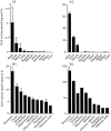
Distribution of N-acyltransferase activity in detergent-solubilized particulate fractions from various tissues (A) and from various brain regions (B) of the rat. N-acyltransferase assays were carried out under standard conditions (see Materials and Methods), using either 1,2-di[14C]arachidonoyl PC (A1, B1) or 1,2-di[14C]palmitoyl PC (A2, B2) as fatty acyl donor. Radioactive N-acyl PEs were purified by monodimensional TLC (A1, A2) or column chromatography (B1, B2) (see Materials and Methods). TLC purification was used in the experiments on tissue distribution because a PLA2 activity was present in some tissues, which produced large quantities of [14C]fatty acids, interfering with the assay by column chromatography (data not shown). Results are expressed as the mean ± SEM of at least three experiments.
Fig. 12.
Fig. 12.
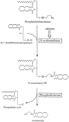
Hypothetical model ofN-arachidonoyl PE biosynthesis in rat brain tissue. Rises in intracellular Ca2+ concentration produced by neuronal activity may stimulate a N-acyltransferase activity, which catalyzes the transfer of arachidonate fromsn-1 arachidonoyl phospholipids (possibly PC) to PE. Ca2+ may also stimulate a D-type phosphodiesterase activity (phospholipase D), which cleaves the distal phosphodiester bond ofN-arachidonoyl PE, forming anandamide. The mass ofN-arachidonoyl PE found in brain is only approximately twofold greater than the mass of anandamide, suggesting that sustained anandamide formation in neurons may require the concomitant stimulation by Ca2+ of N-acyltransferase and phosphodiesterase activities.

References

    1. Aveldano de Caldironi MI, Bazan NG. α-Methyl-p-tyrosine inhibits the production of free arachidonic acid and diacylglycerols in brain after a single electroconvulsive shock. Neurochem Res. 1979;4:213–221. - PubMed
    1. Blank ML, Cress EA, Robinson M, Snyder F. Metabolism of unique diarachidonoyl and linoleoylarachidonoyl species of ethanolamine and choline phosphoglycerides in rat testes. Biochim Biophys Acta. 1985;833:366–371. - PubMed
    1. Cadas H, Gaillet S, Beltramo M, Venance L, Piomelli D. Biosynthesis of an endogenous cannabinoid precursor in neurons and its control by calcium and cAMP. J Neurosci. 1996a;16:3934–3942. - PMC - PubMed
    1. Cadas H, Schinelli S, Piomelli D. Membrane localization of N-acylphosphatidylethanolamine in central neurons: studies with exogenous phospholipases. J Lipid Med Cell Signal. 1996b;14:63–70. - PubMed
    1. Chilton FH, Murphy RC. Stimulated production and natural occurrence of 1,2-diarachidonoylglycerophosphorylcholine in human neutrophils. Biochem Biophys Res Commun. 1987;145:1126–1133. - PubMed

Publication types