. 1997 Feb 15;17(4):1350-62.
doi: 10.1523/JNEUROSCI.17-04-01350.1997.

Ligand-gated ion channel subunit partnerships: GABAA receptor alpha6 subunit gene inactivation inhibits delta subunit expression

Affiliations

Ligand-gated ion channel subunit partnerships: GABAA receptor alpha6 subunit gene inactivation inhibits delta subunit expression

A Jones et al. J Neurosci. .

Abstract

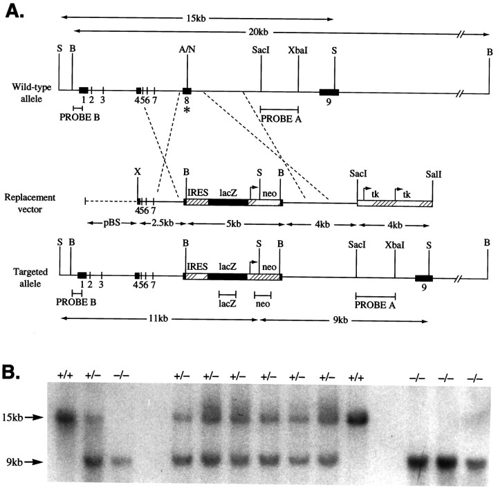
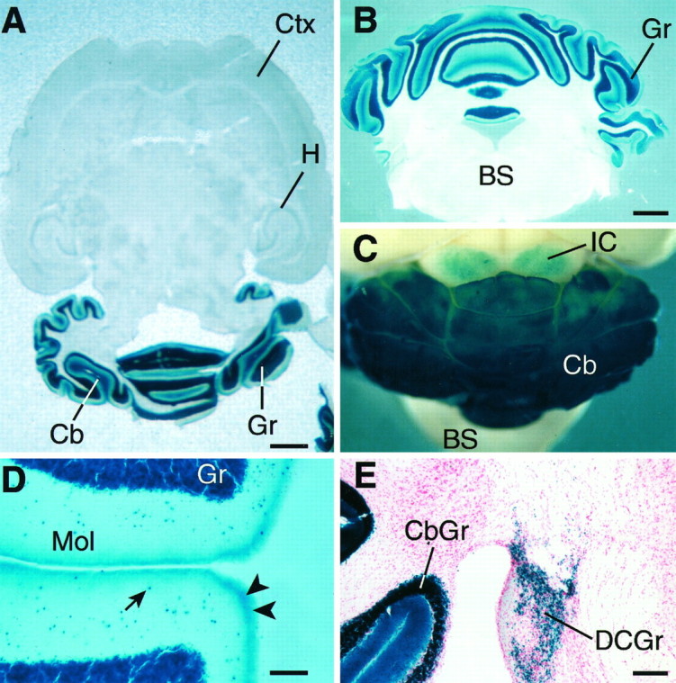
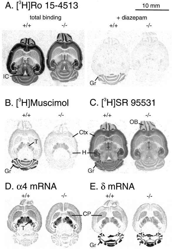
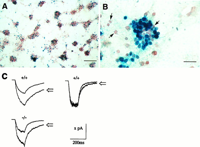
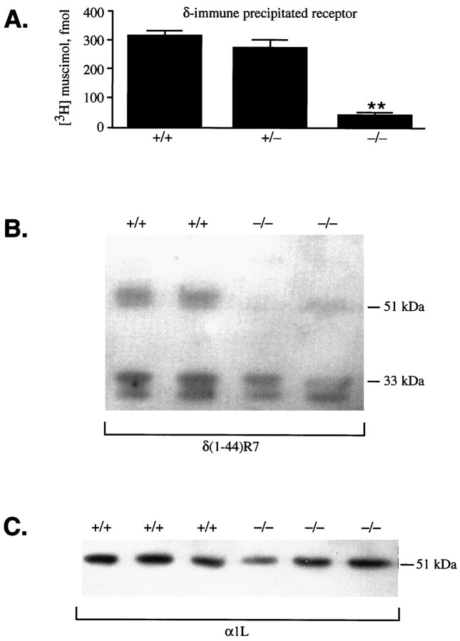
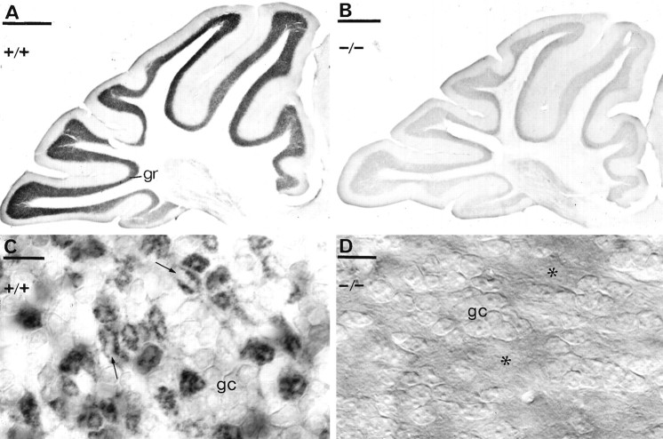
Cerebellar granule cells express six GABAA receptor subunits abundantly (alpha1, alpha6, beta2, beta3, gamma2, and delta) and assemble various pentameric receptor subtypes with unknown subunit compositions; however, the rules guiding receptor subunit assembly are unclear. Here, removal of intact alpha6 protein from cerebellar granule cells allowed perturbations in other subunit levels to be studied. Exon 8 of the mouse alpha6 subunit gene was disrupted by homologous recombination. In alpha6 -/- granule cells, the delta subunit was selectively degraded as seen by immunoprecipitation, immunocytochemistry, and immunoblot analysis with delta subunit-specific antibodies. The delta subunit mRNA was present at wild-type levels in the mutant granule cells, indicating a post-translational loss of the delta subunit. These results provide genetic evidence for a specific association between the alpha6 and delta subunits. Because in alpha6 -/- neurons the remaining alpha1, beta2/3, and gamma2 subunits cannot rescue the delta subunit, certain potential subunit combinations may not be found in wild-type cells.


Figures

Fig. 1.
Fig. 1.
GABAA receptor α6subunit gene disruption by homologous recombination. A, Wild-type α6 gene, targeting (replacement) vector, and the disrupted α6 gene structures. Numbersindicate exons. On the Replacement vector, thebroken line indicates pBluescript (pBS) sequences. On the wild-type allele, theasterisk marks exon 8 where the IRES lacZ/neo cassette was inserted. Only relevant restriction sites are shown. A, AflII;B, BamHI; N,NcoI; S, SphI;X, XhoI. Arrows mark theneo and tk gene promoter sites and direction of transcription. The lacZ coding sequence orientation is the same as the α6 gene, thus permitting its translation from the IRES sequence (striped box) to be initiated on the mRNA derived from the α6 promoter. Expected restriction fragment lengths diagnostic for homologous recombination and the probes used to detect these are marked by double-headed arrows andhorizontal bars, respectively. B, Confirmation of α6 mutant allele germline transmission. Biopsy tail DNA samples were digested with SphI, electrophoresed, and Southern-blotted. The membrane was probed withPROBE A (3′ flanking). Wild-type (+/+) individuals give a 15 kb band, the homozygous null (−/−) animals give a 9 kb band, and heterozygotes (+/−) give both bands.
Fig. 3.
Fig. 3.
LacZ expression driven from the α6gene locus in α6 −/− adult mouse brains illustrated in horizontal (A), coronal (B), and whole-mount views (C); blue colorationindicates lacZ activity. A and B show the confined expression of the α6 gene to the cerebellar granule cell layer; C shows the expression in the dorsal regions of the inferior colliculi; D shows higher-power view of α6 gene expression in the molecular layer of the cerebellum. The arrow indicates an example of the numerous lacZ positive cells in the molecular layer. These are probably nonmigrated granule cells. The arrowheads mark putative parallel fiber staining; E, α6 gene expression in the dorsal cochlear nucleus granule cells.BS, Brainstem; Cb, cerebellum;CbGr, cerebellar granule cell layer; Ctx, neocortex; Gr, cerebellar granule cells;DCGr, dorsal cochlear nucleus granule cells;H, hippocampus; IC, inferior colliculi;Mol, cerebellar molecular layer. Scale bars:A, 1.3 mm; B, C, 1 mm;D, 150 μm; E, 300 μm.
Fig. 2.
Fig. 2.
Immunodetection of the α6 subunit of the GABAA receptor in α6 +/+ (A, C) or α6 −/− (B, C) cerebella as visualized with either light microscopic immunoperoxidase reactions (A, B) or immunoblotting (C). A, B, An intense immunoreactivity for the α6subunits in the granule cell layer (gr) disappeared in α6 −/− mice (B). The sections and one immunoblot were reacted with the same N-terminal domain-specific antibody. Scale bars: A, B, 500 μm.C, The 57 kDa α6 protein is absent in α6 −/− cerebella, as shown with either α6-N, an N-terminal-specific antibody, or α6-C, a C-terminal-specific antibody to the α6 subunit.
Fig. 4.
Fig. 4.
Autoradiographic analysis of GABAAreceptor binding sites in wild-type (+/+) and α6 −/− mice. A, Benzodiazepine sites labeled by 5 nm [3H]R0 15-4513 showing total and diazepam-insensitive binding in the presence of 100 μmdiazepam. B, GABAA receptor sites labeled by 20 nm [3H]muscimol, showing total binding. The nonspecific signal in the presence of 100 μm GABA was at the film background level. C, GABAAreceptor sites labeled by 20 nm [3H]SR 95531, showing total binding. The nonspecific binding signal in the presence of 100 μm GABA was similar in wild-type and −/− brains (data not shown). Similar distinct pharmacological profiles were observed between the wild-type and α6 −/− brains in each of seven pairs of adult mice studied. D, E,In situ hybridization x-ray film autoradiographs of adult mouse brains hybridized with α4 (D) and δ-specific (E) 35S-labeled oligonucleotide probes. Wild-type (+/+) brains are on theleft; α6 −/− brains are on theright. No differences can be seen in subunit mRNA levels between +/+ and −/− brains. Note also the very similar pattern of α4 and δ gene expression in the forebrain/thalamus regions, and the correlation with the distribution of [3H]muscimol (B). Cbgr, Cerebellar granule cells; CP, caudate-putamen;Ctx, cerebral cortex; Gr, cerebellar granule cell layer; H, hippocampus; IC, inferior colliculus; OB, olfactory bulb;T, thalamus.
Fig. 5.
Fig. 5.
Electrophysiological characterization of GABAA receptors in cerebellar granule cells from wild-type and α6 −/− cells. Photomicrographs in Aand B show typical examples of lacZ-expressing cerebellar granule cells, isolated from P5 α6 −/− mouse cerebella, and cultured for 3 weeks. A is a low-magnification view, showing the mosaic of blue(lacZ-positive) cells scattered throughout the culture. Both isolated and clustered blue cells can be seen. Within any given cluster, not all the cells are blue and therefore are not expressing the α6 gene. The cells have been counterstained with neutral red. All electrophysiological recordings were from isolated cells. Arrows inB show examples of non-lacZ-expressing cells. Scale bars: A, 200 μm; B, 30 μm.C, Example responses to 120 msec applications of 20 μm GABA alone, and 20 μm GABA with 1 μm flunitrazepam (open arrows). Thetop row shows an example of wild-type cells (+/+) with GABAA receptors that responded to flunitrazepam (left trace) or were insensitive to flunitrazepam (right trace). The bottom row shows a typical GABAA response from an α6 −/− cell and the associated flunitrazepam potentiation. From left to right and top to bottom, the valuex on the scale bar corresponds to 200, 230, and 170 pA, respectively. The traces were averages of three to five consecutive records.
Fig. 6.
Fig. 6.
Immunoprecipitation and immunoblot analysis of GABAA receptor δ subunit levels in wild-type and α6 −/− cerebella. A, After cerebellar GABAA receptors were solubilized in Triton X-100/deoxycholate, the number of [3H]muscimol binding sites immunoprecipitated by the δ(318–400) antiserum from +/+, +/−, and −/− cerebella was determined (n = 10–14).B, Immunoblot analysis: the marked δ subunit reduction in α6 −/− cerebella detected with the δ(1–44)R7 antiserum. The identity of the low molecular weight doublet (33 and 31 kDa) seen in all samples is unknown. C, A 51 kDa α1 immunoreactive band is present in both −/− and +/+ cerebellar samples as detected with the α1(328–382)/α1L antibody.
Fig. 7.
Fig. 7.
Immunodetection of the δ subunit of the GABAA receptor in α6 +/+ (A,C) or α6 −/− (B, D) cerebella, using a polyclonal antibody δ R7 and immunoperoxidase reaction. The granule cell layer showed intense immunoreactivity in α6 +/+ animals but almost no staining was observed in the α6 −/− mouse. C, At higher magnification, it is evident that the δ subunit is localized mainly in the glomeruli, granule cell bodies (gc) being only weakly outlined. The glomeruli appear as dark rings of granule cell dendrites surrounding a pale center (arrows) representing the unstained mossy fiber terminal. D, In the α6 −/− mice, both the granule cell bodies (gc) and the glomeruli (asterisks) are immunonegative for the δ subunit. C andD were photographed using DIC optics. Scale bars:A, B, 500 μm; C, D, 10 μm.

References

    1. Bahn S, Harvey RJ, Darlison MG, Wisden W. Conservation of γ aminobutyric acid type A receptor α6 subunit gene expression in cerebellar granule cells. J Neurochem. 1996;66:1810–1818. - PubMed
    1. Benke D, Mertens S, Trzecia A, Gillessen D, Möhler H. Identification and immunohistochemical mapping of GABAA receptor subtypes containing the δ subunit in rat brain. FEBS Lett. 1991;283:145–149. - PubMed
    1. Bonnerot C, Nicolas J-F. Application of LacZ gene fusions to postimplantation development. Methods Enzymol. 1993;225:451–469. - PubMed
    1. Caruncho HJ, Costa E. Double-immunolabelling study of GABAA receptor subunits in label-fracture replicas of cultured rat cerebellar granule cells. Receptors Channels. 1994;2:143–153. - PubMed
    1. Caruncho HJ, Puia G, Möhler H, Costa E. The density and distribution of six GABAA receptor subunits in primary cultures of rat cerebellar granule cells. Neuroscience. 1995;67:583–593. - PubMed

Publication types

MeSH terms

Associated data