. 1997 Feb 15;17(4):1425-34.
doi: 10.1523/JNEUROSCI.17-04-01425.1997.

The expression and function of Notch pathway genes in the developing rat eye

Affiliations

The expression and function of Notch pathway genes in the developing rat eye

Z Z Bao et al. J Neurosci. .

Abstract

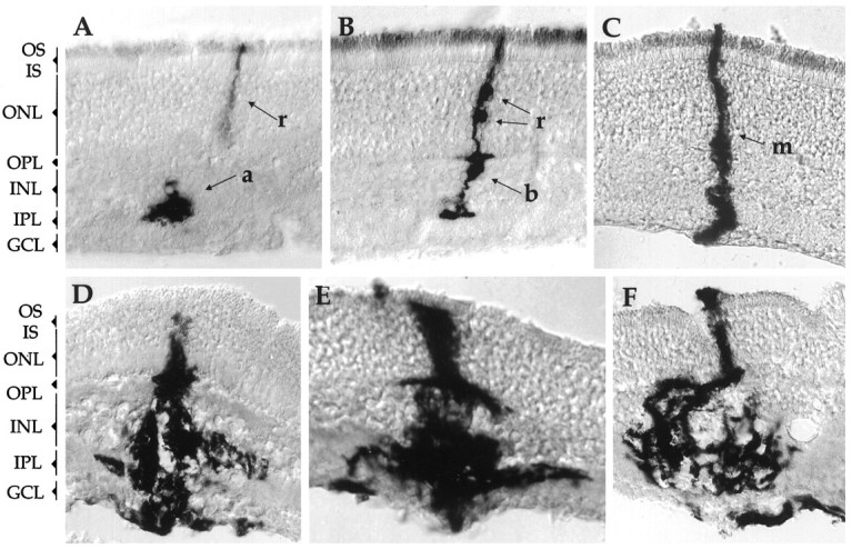
The Notch gene plays a role in the development of disparate tissues in multiple organisms. Because the vertebrate eye is an excellent model system for both patterning and cell fate determination, two processes that can involve Notch, we examined the expression patterns of Notch 1 and Notch 2, and their ligands Delta and Jagged, in the developing rat eye. Notch 1 and Delta were found to be expressed in the neural retina during the period of cell fate determination and differentiation. Notch 2 was found to be expressed in the non-neuronal derivatives of the optic cup, including the pigment epithelium, optic stalk, and ciliary body. Jagged was expressed in distinct regions within the optic vesicle, ciliary body, and lens, with patterns that changed over time. The potential function of Notch 1 in cell-type specification and differentiation was examined by introducing a constitutively active form of Notch 1 in vivo using a replication-incompetent retrovirus. This form of Notch 1 was found to cause abnormal growth and interfere with the differentiation of multiple retinal cell types.


Figures

Fig. 5.
Fig. 5.
Viral constructs for expressing truncated forms of the Notch 1 gene. A, Full-length Notch 1 protein consists of extracellular EGF repeats, Lin/Notch repeats (LN), transmembrane domain (TM), cytoplasmic cdc10/ankyrin repeats, and aPEST sequence. Truncations of Notch, mNIC, and mNEC, with myc epitope tags, were inserted into the cloning site of pLIA.B, pLIA retroviral vector. On the basis of the MMLV backbone, pLIA has a cloning site for expressing exogenous genes under the transcriptional control of the LTR. It also contains an IRES sequence directing the translation of a marker gene, PLAP.
Fig. 1.
Fig. 1.
Expression of Delta andJagged in the optic vesicle at E12.5. A, Diagram of the E12.5 optic vesicle. B, In situ hybridization of a section with the Deltaprobe. Delta was not detected in the optic vesicle region (marked by arrowheads). In contrast,Delta hybridization was observed in a subset of cells in the VZ of the forebrain (F). C,In situ hybridization with the Jaggedprobe. Jagged hybridization was observed in the dorsal region of both PNR and lens placode. It was also expressed in the VZ of the forebrain. D, Whole-mount in situhybridization with the Jagged probe.Jagged hybridization was observed in the dorsal region of the retina (arrow) and lens placode (arrowhead). LP, Lens placode;PPE, presumptive pigment epithelium; PNR, presumptive neural retina.
Fig. 2.
Fig. 2.
Expression of Notch 1, Notch 2, Delta, and Jagged in the E15.5 eye cup. Coronal sections of an E15.5 rat eye (ventral side) are shown.A, Notch 1 expression was observed only in the neural retina. B, Notch 2expression was observed in the PE and optic stalk. C,Delta expression was observed in neural retina, similar to that of Notch 1. D,Jagged hybridization signal was observed in the presumptive ciliary body and the equatorial region and anterior of the lens. In contrast to the earlier dorsal expression patterns, the expression of Jagged was largely symmetrical in the dorsal and ventral sides of the optic cup and lens (inset). NR, Neural retina;CB, ciliary body region; I, iris;L, lens; OS, optic stalk;PE, pigment epithelium.
Fig. 3.
Fig. 3.
Expression of Notch 1, Notch 2, Delta, and Jagged in the postnatal eye. A, Diagram of the cellular composition of the P5 retina. At this age, three layers can be distinguished. The ganglion cell layer contains mostly differentiated ganglion cells; the inner plexiform layer consists of dendritic processes of the differentiated amacrine cells and ganglion cells; the VZ has both differentiated and undifferentiated cells at this stage. Differentiated cone photoreceptors and some rod photoreceptors are located within the top half of the VZ, whereas horizontal cells are within the center and bottom half of the VZ. B,Notch 1 in P5 retina; C, Notch 1 in adult retina; D, Delta in P5 retina; E, Notch 2 in P5 eye;F, Jagged in P5 eye. Note that bothNotch 2 and Jagged are expressed in the ciliary body (arrows). A subset of cells in the INL and GCL also express Jagged (arrowheads).GCL, Ganglion cell layer; IPL, inner plexiform layer; VZ, ventricular layer:N, undifferentiated neuroblast; c, cone photoreceptor; h, horizontal cell; a, amacrine cell; g, ganglion cell; r, rod photoreceptor.
Fig. 4.
Fig. 4.
Schematic summary of Notch 1,Notch 2, Delta, and Jaggedexpression patterns in the developing eye, at E12.5 (A, B), E15.5 (C, D), P0 (E, F). Notch 1 and Jaggedpatterns are shown in A, C, E; Notch 2and Delta patterns are summarized in B, D, F.
Fig. 6.
Fig. 6.
Expression of the truncated Notch 1gene in infected 3T3 cells, detected by immunohistochemical staining with an anti-myc antibody, 9E10. A, mNIC-infected 3T3 cells stained with the 9E10 monoclonal antibody (mAb).B, mNIC-infected 3T3 cells stained with X-Phos/NBT shows the expression of PLAP. C, mNEC-infected cells stained with the 9E10 mAb. Note that the Notch 1 intracellular domain was mostly translocated to nuclei, whereas the Notch 1 extracellular domain was localized to the cell membrane.
Fig. 7.
Fig. 7.
Clonal morphology of LIA- and mNIC-infected clones. P0 retinae were infected in vivo by intraocular injection. Retinae were harvested 3 weeks later, stained with X-Phos/NBT, and then sectioned to reveal clonal morphology.A–C, Control LIA-infected clones. Four cell types were observed: rod photoreceptor, amacrine cell, bipolar cell, and Müller glia. D–F, mNIC-infected clones. Note the grossly abnormal morphology within clones infected with the virus expressing an activated form of Notch 1. OS, Outer segment layer; IS, inner segment layer;ONL, outer nuclear layer; OPL, outer plexiform layer; INL, inner nuclear layer;IPL, inner plexiform layer; GCL, ganglion cell layer; a, amacrine cell; r, rod photoreceptor; b, bipolar cell; m, Müller glia.
Fig. 8.
Fig. 8.
Morphology of mNIC- and mNEC-infected clones within retinal explants. E18 retinal explants were co-infected with BAG virus, encoding lacZ, and mNIC virus (A) or mNEC virus (B). The explants were harvested 2 weeks later and processed to visualize both BAG-infected clones (blue) and mNIC- or mNEC-infected clones (purple). Photographs were taken from the photoreceptor side of the retina. Note mNIC-infected clones were very large, with abnormal morphology, including extensive processes. In contrast, mNEC-infected clones appeared normal. The effect of the activated Notch appeared to be restricted to infected cells, because blue clones were normal even when located very close to mNIC-infected clones (arrows).

References

    1. Ahmed I, Zagouras P, Artavanis-Tsakonas S. Involvement of Notch-1 in mammalian retinal neurogenesis: association of Notch-1 activity with both immature and terminally differentiated cells. Mech Dev. 1995;53:73–85. - PubMed
    1. Alexiades MR, Cepko C. Quantitative analysis of proliferation and cell cycle length during development of the rat retina. Dev Dyn. 1996;205:293–307. - PubMed
    1. Artavanis-Tsakonas S, Matsuno K, Fortini ME. Notch signaling. Science. 1995;268:225–232. - PubMed
    1. Austin CP, Feldman DE, Ida JA, Jr, Cepko CL. Ganglion cells in the vertebrate retina are selected from an equivalence group regulated by Notch. Development. 1995;121:3637–3650. - PubMed
    1. Ausubel FM, Brent R, Kingston RE, Moore DD, Seidman JG, Smith JA, Struhl K (1996) Current protocols in molecular biology. New York: Greene/Wiley-Interscience.

Publication types

MeSH terms