. 1997 Feb 15;17(4):1471-80.
doi: 10.1523/JNEUROSCI.17-04-01471.1997.

Phospholipase C-beta1 is present in the botrysome, an intermediate compartment-like organelle, and Is regulated by visual experience in cat visual cortex

Affiliations

Phospholipase C-beta1 is present in the botrysome, an intermediate compartment-like organelle, and Is regulated by visual experience in cat visual cortex

P C Kind et al. J Neurosci. .

Abstract

Monoclonal antibody Cat-307 identifies a 165 kDa neuronal protein expressed in the cat visual cortex during the period of sensitivity to alterations in visual experience (). Dark-rearing, which prolongs the sensitive period, also prolongs the expression of the Cat-307 protein. The Cat-307 protein localizes to an organelle, here called the botrysome (from the Greek botrys, cluster of grapes), that is located between the endoplasmic reticulum (ER) and Golgi apparatus. The botrysome is composed of small ring-shaped profiles with electron-dense coats. The size and morphology of the rings and their coats are similar to those described for ER to Golgi transport vesicles. Biochemically, the Cat-307 protein cofractionates with microsomes and partitions with subunits of the coatomer proteins that coat ER-to-Golgi transport vesicles. Partial amino acid sequencing reveals that the Cat-307 protein is phospholipase C-beta1, the G-protein-dependent phosphodiesterase that hydrolyses phosphatidylinositol 4,5 biphosphate into inositol 1,4,5 triphosphate and diacylglycerol after the stimulation of a variety of neurotransmitter receptors at the cell surface. These results suggest a role for phospholipase C-beta1 and the botrysome in developmental plasticity and provide a possible link between receptor activation at the cell surface and protein transport during neuronal development.


Figures

Fig. 6.
Fig. 6.
The Cat-307 protein cofractionates with microsomal membranes and with β-COP. A crude microsomal fraction prepared by differential centrifugation (Malhotra et al., 1989) contains the Cat-307 protein (A, lane S). The microsomal fraction was then further purified in a step sucrose gradient (A, lanes 1-10,B). A, Western blot analysis shows that the protein recognized by Cat-307 comigrates on a step sucrose density gradient with microsomes (no triton, fractions 5 and 6). Little immunoreactivity is detected at the bottom of the gradient (B, no triton). However, after treatment of the microsomes with Triton X-100 before centrifugation, the Cat-307 protein is retained in the bottom fraction of the gradient (B,triton). Triton X-100 solubilizes microsomal membranes; thus, the lack of floatation of the Cat-307 protein in the presence of Triton X-100 indicates that the floatation in the gradient in the absence of Triton X-100 is attributable to an association of the protein with intact microsomal membranes. B, Western blot analyses of β-COP shows an identical fractionation pattern to that observed for the Cat-307 protein; that is, in the absence of Triton X-100, immunoreactivity for β-COP is seen in fractions 5 and 6, thereby confirming the association of the Cat-307 protein with the microsomal membrane fractions. Furthermore, pretreatment of the microsomal membranes with 1% Triton X-100 prevents the floatation of β-COP in the sucrose density gradient; thus, immunoreactivity for β-COP is no longer detected in fractions 5 and 6 but is seen exclusively at the bottom of the gradient.
Fig. 1.
Fig. 1.
Dark-rearing prevents the normal downregulation of the Cat-307 protein in primary visual, but not in primary auditory, cortex. Sections through layers IV and V of primary visual (A, B, C) and primary auditory (D, E, F) cortex from a 5-week-old normally reared (A,D), a 15-week-old normally reared (B,E), and a 17-week-old dark-reared (C,F) cat immunoreacted with Cat-307. Immunoreactive botrysomes are present in both the visual (A) and the auditory (D) cortices in the 5-week-old cat but absent from both areas by 15 weeks of age (B,E). Botrysomes are present in the visual cortex (C) after dark-rearing, but are not present in the auditory cortex (F) of dark-reared animals. Immunoreactive botrysomes can be found in all layers of area 17 of dark-reared animals in a pattern that is very similar to that observed in 5-week-old normal animals. Scale bar, 30 μm.
Fig. 2.
Fig. 2.
Cat-307 identifies a cytoplasmic structure in neurons that is often located near a major dendrite.AC, Three layer V neurons containing Cat-307 immunoreactive botrysomes. In 50-μm-thick sections, Cat-307 immunoreactive profiles are observed in the cytoplasm of neurons and are most often found within, or just below, the apical dendrites of pyramidal cells. A, Two botrysomes, one located at the base of the large apical dendrite of a layer V pyramidal neuron (arrow) and the other in the basal portion of a neuron (arrowhead). The apical dendrite of the pyramidal neuron (arrow) contains a small botrysome farther from the cell body. B, A layer V pyramidal cell with a botrysome located just below the apical dendrite and very close to the nucleus.C, A large layer V pyramidal cell containing three immunoreactive botrysomes within its cell body and apical dendrite. Two of the botrysomes are associated with the apical dendrite (arrows), one located within and the other at the base of the dendrite. The third is located in the basal portion of the cell body (arrowhead). Such a basally located botrysome would have been classified as not associated with a dendrite; however, the basal dendrites of the cell are out of the plane of section, and this botrysome may also be at the base of a dendrite. The largest botrysomes are seen in layer V neurons, where they can reach a diameter of 4 μm. The diameter of botrysomes varies; even within a single cell, botrysome size does not correlate with neuronal size, although the smallest botrysomes are usually found within dendrites. Scale bar, 20 μm.
Fig. 3.
Fig. 3.
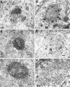
Cat-307 identifies a neuronal organelle, the botrysome, that is located between the ER and thecis-Golgi and that can be identified independently of Cat-307 immunoreactivity. A, B, Electron micrographs showing two examples of Cat-307 immunoreactivity in botrysomes located between the ER (ER) and thecis-Golgi (cG). Cat-307 immunoreactivity has never been observed within the Golgi stack (GS) or on the trans face of the Golgi (tG). The immunoreactive botrysome (arrowheads) is a compact, ovoid structure that is a conglomeration of small, ring-shaped profiles. The DAB reaction product, however, prevents a detailed analysis of the substructure of the organelle. Scale bars: A,B, 350 nm. C, Another example of an immunoreactive botrysome (large arrowheads) showing that the internal structure is not uniform (see also B). The center of the botrysome has a dense core (small arrowheads) with less dense flanking regions (asterisks). The ring-shaped elements surround the core and are the major components of the botrysome. Although the example inC does not demonstrate an association with the Golgi complex, three-dimensional reconstructions of single botrysomes suggest that all botrysomes are associated with the Golgi complex. Scale bar, 350 nm. D, Electron micrographs of Cat-307 immunoreactivity visualized with a gold-conjugated secondary antibody enhanced with silver demonstrate that the immunoreactive botrysome (arrowheads) is an organelle that can be identified independent of HRP histochemistry. Scale bar, 140 nm. E,F, Low- (E) and high-power (F) electron micrographs of a botrysome (arrowheads in E) that was deep in the section, inaccessible to the gold-conjugated antibody. (In tissue in which an HRP-conjugated secondary antibody was used, we have never seen an unlabeled botrysome.) The low-power micrograph (E) clearly demonstrates the location of the botrysome between the Golgi apparatus and the ER. Higher magnification (F) of this unlabeled botrysome confirms it to be a conglomeration of small ring-shaped profiles. The diameter of an individual ring is ∼70 nm, similar to the size of COP-coated vesicles. The rings have a moderately electron dense wall with a clear center. Not all of the rings are complete; many are c-shaped. The rings are covered with an electron-dense material with a thickness of 15 nm, similar to the protein coat of transport vesicles between the ER and Golgi apparatus. Scale bars: E, 650 nm; F, 100 nm.
Fig. 4.
Fig. 4.
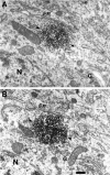
The botrysome is always associated with the ER and Golgi complex. Sections from a serial reconstruction of a single botrysome (arrowheads). In A, the botrysome is not closely associated with the Golgi complex (G); however, the micrograph in B of a subsequent section clearly demonstrates the close approximation of the botrysome and the Golgi apparatus. Scale bar, 200 nm. We have partially reconstructed four botrysomes, all of which were associated with both the ER (ER) and Golgi apparatus. Scale bar, 800 nm.
Fig. 5.
Fig. 5.
The Golgi apparatus is not always associated with the botrysome. Low-power electron micrograph of three neurons (outlined in dotted lines) in layer V of kitten visual cortex. One neuron contains a Cat-307 immunoreactive botrysome (arrowheads) labeled with HRP, located adjacent to a Golgi apparatus (G). The adjacent neuron contains numerous Golgi apparati but no botrysomes. Golgi apparati are much more numerous than botrysomes and can be seen throughout the cell body. The botrysome appears to be associated with a specialized region of the Golgi apparatus located near the base of dendrites. Scale bar, 3 μm.
Fig. 7.
Fig. 7.
The Cat-307 protein is PLC-β1. A, Cortical tissue was homogenized in either 320 mm sucrose (lanes 1-5) or PBS (lanes 6–10). The homogenates (H, lanes 1 and 6) were spun at 100,000 ×g for 1 hr and separated into supernatant (S1, lanes 2 and 7) and pellet fractions. The pellet fraction was washed in an equal volume of the appropriate buffer and spun at 100,000 × g. The supernatant fraction was removed (W, lanes 3 and 8) and the pellet rehomogenized in an equal volume of PBS. The H was allowed to sit for 20–30 min at 4°C and was then centrifuged for 1 hr. The supernatant (S2,lanes 4 and 9) and pellet (P2, lanes 5 and 10) fractions were collected, and all fractions were subjected to Western blot analysis for the Cat-307 protein. The large amount of Cat-307 protein in the S2 fraction of the sucrose homogenates compared with the PBS homogenates indicates that the Cat-307 protein is associated with the microsomal membranes in a noncovalent manner that is dependent on the osmolarity of the homogenizing solution. This type of membrane association is characteristic of peripheral membrane proteins and is identical to that shown previously for the COPs. The Cat-307 protein was further concentrated and purified from the S2 fraction by ammonium sulfate precipitation and FPLC using a monoQ column (data not shown). Amino acid sequence analysis identified the Cat-307 protein as PLC-β1. B, Immunoblot of purified Cat-307 protein stained with antibodies to Cat-307 (lane 1) or PLC-β1 (lane 2). One lane of the purified protein was transferred to nitrocellulose, cut in half, and stained for the two antibodies. Cat-307 and antibody to PLC-β1 each identifies an exactly comigrating protein doublet. Proteins immunoprecipitated with either PLC-β1 (lanes 4 and 6) or Cat-307 (lanes 3 and 5) are immunoreactive for both Cat-307 (lanes 5 and6) and PLC-β1 (lanes 3 and4). Each antibody immunoprecipitates a protein recognized by the other antibody, indicating that the Cat-307 protein is PLC-β1.

Similar articles

Cited by

References

    1. Antonini A, Stryker MP. Rapid remodeling of axonal arbors in the visual cortex. Science. 1993a;260:1819–1821. - PubMed
    1. Antonini A, Stryker MP. Development of individual geniculocortical arbors in cat striate cortex and effects of binocular impulse blockade. J Neurosci. 1993b;13:3549–3573. - PMC - PubMed
    1. Bashir ZI, Collingridge GL. Synaptic plasticity: long-term potentiation in the hippocampus. Curr Opin Neurobiol. 1992;2:328–335. - PubMed
    1. Bashir ZI, Bortolotto ZA, Davies CH, Berretta N, Irving AJ, Seal AJ, Henley JM, Jane DE, Watkins JC, Collingridge GL. Induction of LTP in the hippocampus needs synaptic activation of glutamate metabotropic receptors. Nature. 1993;363:347–350. - PubMed
    1. Bauerfiend FR, Huttner WB. Biogenesis of constitutive secretory vesicles, secretory granules and synaptic vesicles. Curr Opin Cell Biol. 1993;5:628–635. - PubMed

Publication types

MeSH terms

LinkOut - more resources