. 1997 Jan 13;136(1):29-43.
doi: 10.1083/jcb.136.1.29.

Chromosomes with two intact axial cores are induced by G2 checkpoint override: evidence that DNA decatenation is not required to template the chromosome structure

Affiliations

Chromosomes with two intact axial cores are induced by G2 checkpoint override: evidence that DNA decatenation is not required to template the chromosome structure

P R Andreassen et al. J Cell Biol. .

Abstract

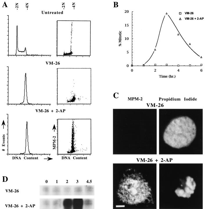
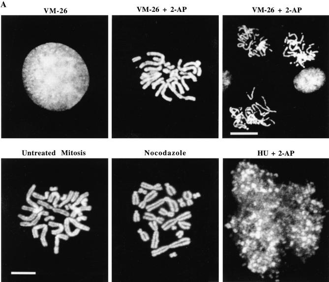
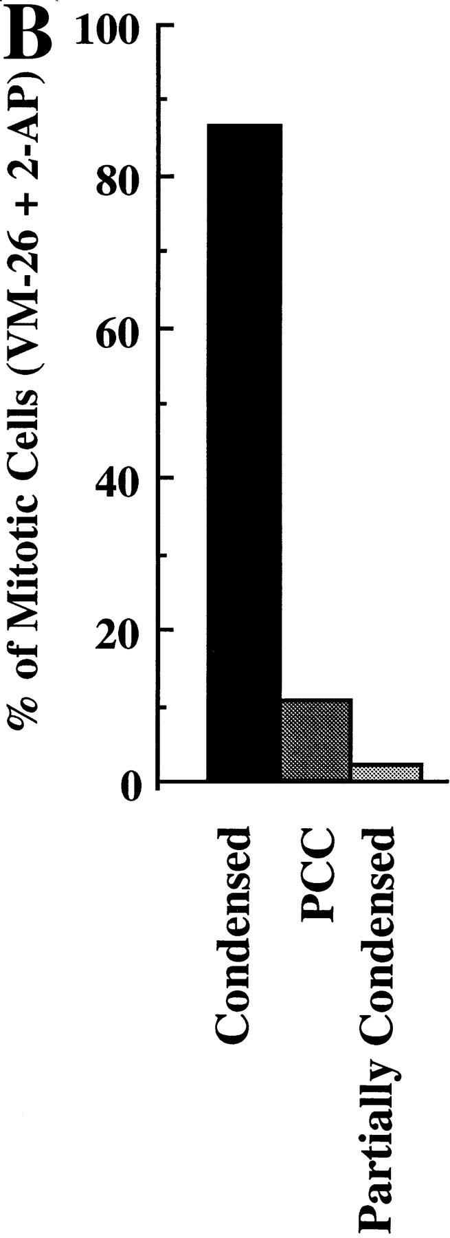
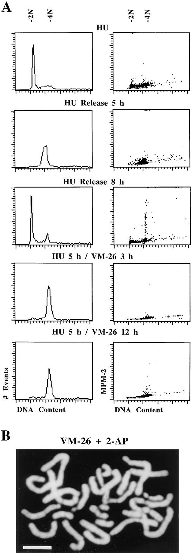
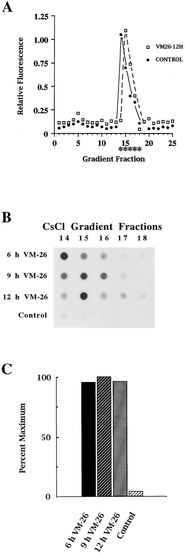
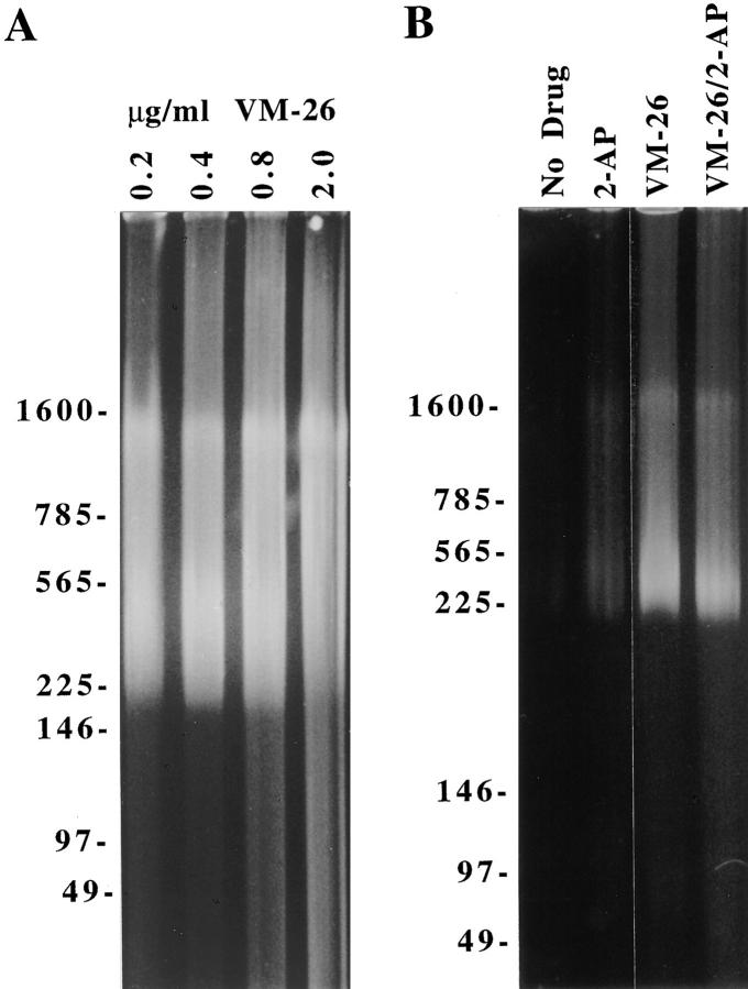
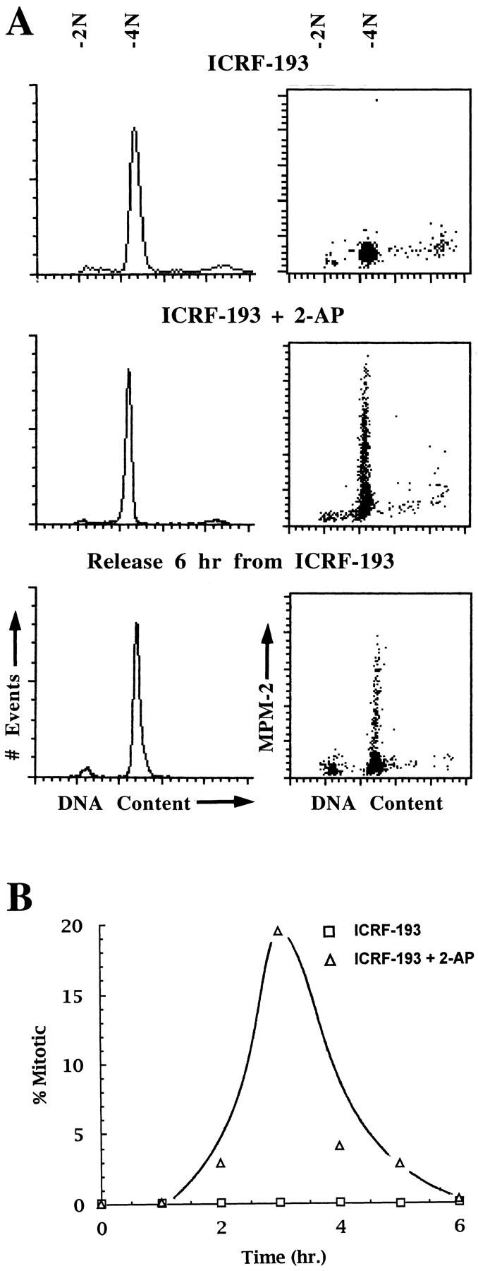
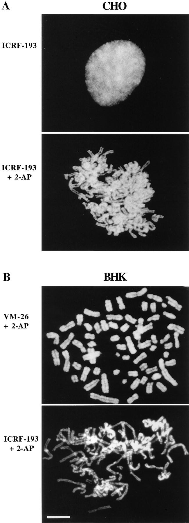
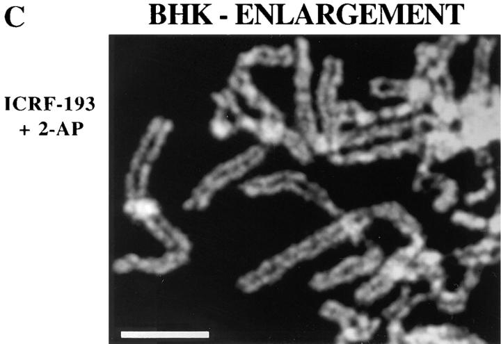
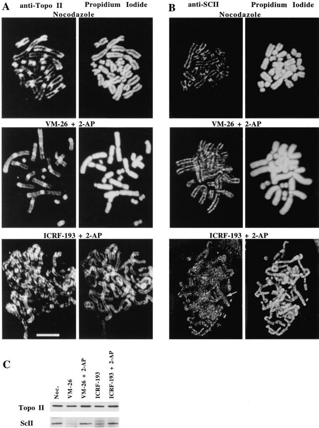
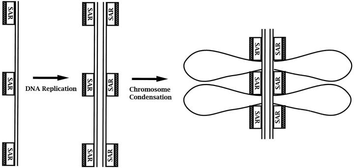
Here we report that DNA decatenation is not a physical requirement for the formation of mammalian chromosomes containing a two-armed chromosome scaffold. 2-aminopurine override of G2 arrest imposed by VM-26 or ICRF-193, which inhibit topoisomerase II (topo II)-dependent DNA decatenation, results in the activation of p34cdc2 kinase and entry into mitosis. After override of a VM-26-dependent checkpoint, morphologically normal compact chromosomes form with paired axial cores containing topo II and ScII. Despite its capacity to form chromosomes of normal appearance, the chromatin remains covalently complexed with topo II at continuous levels during G2 arrest with VM-26. Override of an ICRF-193 block, which inhibits topo II-dependent decatenation at an earlier step than VM-26, also generates chromosomes with two distinct, but elongated, parallel arms containing topo II and ScII. These data demonstrate that DNA decatenation is required to pass a G2 checkpoint, but not to restructure chromatin for chromosome formation. We propose that the chromosome core structure is templated during interphase, before DNA decatenation, and that condensation of the two-armed chromosome scaffold can therefore occur independently of the formation of two intact and separate DNA helices.


Figures

Figure 1
Figure 1
2-AP overrides VM-26–induced G2 arrest in CHO cells. (A) Histograms of DNA content (left) and two-dimensional dot plots of MPM-2, a mitosis-specific phosphoepitope, versus DNA content (right). In comparison to untreated controls, 16 h of treatment with 0.20 μg/ml VM-26 results in accumulation of cells in G2 with 4N DNA content and low MPM-2 signal. Addition of 10 mM 2-AP during continuous exposure to VM-26 induces entry into mitosis, as indicated by the accumulation of cells with an elevated MPM-2 signal. (B) Quantitation of mitotic entry induced by 2-AP. Quantitation was performed by microscopy, counting cells that were both positive for MPM-2 and for chromatin condensation, as visualized with propidium iodide. Cells were synchronized in G2 by 16 h of exposure to VM26 and were subsequently exposed to both VM-26 and 2-AP or were continued in VM-26 alone. All points represent the average of three counts of at least 250 cells each. Standard deviation was <1.2% of the ordinate scale for each value. (C) Micrographs showing the MPM-2 immunofluorescence image of cells treated with VM-26 (top) or with both VM-26 and 2-AP (bottom). MPM-2 levels are low in interphase cells, which are identified by the presence of propidium iodide stained nuclei (right). In contrast, MPM-2 levels are elevated in mitotic cells, also containing condensed chromatin, after induction by 2-AP. (D) Assay of p34cdc2 activity in cells arrested in G2 with VM-26, and in VM-26–treated cells induced to enter mitosis with 2-AP. Cells were pretreated with VM-26 for 16 h before the time points of the assay. Bar, 5 μm.
Figure 2
Figure 2
Condensed chromosomes form when CHO cells, blocked in G2 with VM-26, are induced to enter mitosis with 2-AP. (A) Chromosome spreads of mitotic cells treated with VM-26 and 2-AP, and spreads from untreated mitotic cells, or from cells arrested in mitosis with 0.20 μg/ml nocodazole. For comparison, images are also shown of VM-26–treated G2 nuclei and of chromosome fragmentation in spreads from cells blocked in S-phase with 2 mM HU and then advanced to mitosis by addition of 2-AP. (B) Quantitation of the percentage of VM-26–blocked cells, treated with 2-AP, that contain condensed chromosomes at mitosis. Counts were made from chromosome spreads prepared by a 16 h block in VM-26, followed by a 3-h treatment with both VM-26 and 2-AP. For comparison, the percentage of mitotic cells with incompletely condensed chromosomes or with fragmented chromosomes (PCC) were also quantitated. All data are derived from three counts each of 150 or more mitotic cells. Bars: (lower left) 10 μm (for all panels except upper right); (upper right) 20 μm.
Figure 2
Figure 2
Condensed chromosomes form when CHO cells, blocked in G2 with VM-26, are induced to enter mitosis with 2-AP. (A) Chromosome spreads of mitotic cells treated with VM-26 and 2-AP, and spreads from untreated mitotic cells, or from cells arrested in mitosis with 0.20 μg/ml nocodazole. For comparison, images are also shown of VM-26–treated G2 nuclei and of chromosome fragmentation in spreads from cells blocked in S-phase with 2 mM HU and then advanced to mitosis by addition of 2-AP. (B) Quantitation of the percentage of VM-26–blocked cells, treated with 2-AP, that contain condensed chromosomes at mitosis. Counts were made from chromosome spreads prepared by a 16 h block in VM-26, followed by a 3-h treatment with both VM-26 and 2-AP. For comparison, the percentage of mitotic cells with incompletely condensed chromosomes or with fragmented chromosomes (PCC) were also quantitated. All data are derived from three counts each of 150 or more mitotic cells. Bars: (lower left) 10 μm (for all panels except upper right); (upper right) 20 μm.
Figure 3
Figure 3
Condensed chromosomes form in CHO cells after 2-AP– induced override of short-term arrest with VM-26. (A) Histograms of DNA content (left) and two-dimensional dot plots of MPM-2 versus DNA content (right). Cells arrested with 2 mM HU were first presynchronized in G1 by growth in isoleucine- deficient medium. Treatment with 0.20 μg/ml VM-26 beginning at 5 h after release from HU results in arrest at G2 with 4N DNA content and low MPM-2 signal. (B) Condensed chromosomes form in synchronous cells treated with VM-26 for 3 h when they are induced to enter mitosis through checkpoint override by addition of 2-AP in the continued presence of VM-26 for 3 h. Cells were released from HU for 5 h and then treated with VM-26 just before arrival at G2. Bar, 10 μm.
Figure 4
Figure 4
VM-26 treatment stably maintains DNA–topo II covalent complexes in G2-arrested cells. The unchanging presence of covalent complexes indicates suppression of topo II activity. Covalent complexes of DNA and topo II were isolated on cesium chloride gradients after lysis of cells in the presence of sarkosyl. (A) The fractions containing DNA were identified by fluorescence measurement at an emission wavelength of 460 nm after Hoechst 33258 staining. DNA was always confined to fractions 14–18 in repeated experiments. The fractions shown in anti–topo II immunoblots are indicated with asterisks. (B) Topo II–DNA complexes were assayed by anti–topo II immunoblots of fractions containing DNA for cells exposed to 0.20 μg/ml VM-26 for 6, 9, or 12 h after release from presynchronization with isoleucine- deficient medium and then HU. A control, representing cells subjected to synchronization but receiving no treatment with VM-26, is also shown. (C) The relative levels of topo II present in complexes with DNA were quantitated by scanning the dot blots in b and integrating and summing the signals displayed by all the fractions containing DNA.
Figure 5
Figure 5
VM-26 inhibits topo II activity both during G2 and at mitosis induced by checkpoint override with 2-AP. Inhibition of topo II activity was assayed by FIGE analysis of DNA fragments formed by VM-26 stabilization of topo II–DNA covalent intermediates. (A) The size and abundance of fragments is equivalent for cells treated for 16 h with 0.20, 0.40, 0.80, or 2.0 μg/ml VM-26 (0.31, 0.61, 1.22, and 3.1 μM VM-26, respectively). (B) The size and abundance of DNA fragments are equivalent, comparing G2 cells, obtained by 16 h of exposure to 0.20 μg/ml VM-26, with mitotic cells collected by selective detachment after 16 h of treatment with VM-26 and then 3 h of exposure to VM-26 and 2-AP. By comparison, fragmentation is minimal in untreated control cells or in cells treated for 3 h with 2-AP alone.
Figure 6
Figure 6
2-AP overrides ICRF-193–induced G2 arrest in CHO cells. (A) Histograms of DNA content (left) and two-dimensional dot plots of MPM-2 versus DNA content (right). Cells treated with ICRF-193 alone for 16 h contain 4N DNA. After 16 h of ICRF-193 treatment, cells enter mitosis when treated with both ICRF-193 and 10 mM 2-AP for 3 h, as indicated by elevated MPM-2 levels in cells with 4N DNA content. The result is equivalent to mitotic entry observed after removal of ICRF-193 for 6 h. (B) Quantitation of mitotic entry induced by 2-AP in ICRF-193– blocked G2 cells. Mitotic cells were counted at the time points indicated by microscopic assay, scoring cells with both elevated MPM-2 levels and condensed chromatin, as detected by propidium iodide stain. Cells were arrested for 16 h in ICRF-193 before addition of 2-AP at the first time point. Each point represents the average of three counts of at least 250 cells each. Standard deviations were always <1.2% of the ordinate scale.
Figure 7
Figure 7
Partially condensed chromosomes containing two paired arms form at mitosis after 2-AP–induced override of ICRF-193 G2 arrest. (A) Chromosome spreads of CHO cells treated with ICRF-193 for 16 h and then for 3 h with both ICRF-193 and 2-AP contain elongated chromosomes with two arms. In contrast, cells treated with ICRF-193 alone contain only nuclei with uncondensed chromatin. (B) Spreads of chromosomes formed by BHK cells arrested in G2 with VM-26 or ICRF-193 induced to enter mitosis by addition of 2-AP. As in CHO cells, VM-26 does not inhibit full chromosome formation and ICRF-193 results in the formation of elongated chromosomes with well-resolved parallel arms. (C) Enlargement of chromosomes from same cell treated with ICRF-193 and 2-AP, shown in part B. Chromosomes are visualized by staining with propidium iodide. Bars: (a and b) 10 μm; (c) 5 μm.
Figure 7
Figure 7
Partially condensed chromosomes containing two paired arms form at mitosis after 2-AP–induced override of ICRF-193 G2 arrest. (A) Chromosome spreads of CHO cells treated with ICRF-193 for 16 h and then for 3 h with both ICRF-193 and 2-AP contain elongated chromosomes with two arms. In contrast, cells treated with ICRF-193 alone contain only nuclei with uncondensed chromatin. (B) Spreads of chromosomes formed by BHK cells arrested in G2 with VM-26 or ICRF-193 induced to enter mitosis by addition of 2-AP. As in CHO cells, VM-26 does not inhibit full chromosome formation and ICRF-193 results in the formation of elongated chromosomes with well-resolved parallel arms. (C) Enlargement of chromosomes from same cell treated with ICRF-193 and 2-AP, shown in part B. Chromosomes are visualized by staining with propidium iodide. Bars: (a and b) 10 μm; (c) 5 μm.
Figure 8
Figure 8
Both topo II and ScII are present in paired axial cores of chromosomes formed by addition of 2-AP after G2 arrest with either VM-26 or ICRF193. (A) Topo II (left) was immunolocalized in chromosome spreads of mitotic cells treated with either nocodazole or both VM-26 and 2-AP, or both ICRF-193 and 2-AP. Chromosomes were visualized in images generated by counterstaining with propidium iodide (right). (B) ScII (left) was immunolocalized in chromosome spreads during mitotic arrest with nocodazole or after 2-AP–induced override of G2 arrest with VM-26 or ICRF-193. Primary rabbit antibodies against topo II and ScII were detected with FITC-conjugated goat anti–rabbit secondary antibodies. (C) Immunoblots of isolated chromosomes and nuclei demonstrate that topo II is present at equivalent levels both during G2 arrest with topo II inhibitors and in mitosis after 2-AP–induced checkpoint override of G2-arrested cells. By contrast, ScII is present in chromatin only in mitosis either in normal mitotic cells or after 2-AP override of G2 arrest induced by inhibitors of topo II. Bar, 10 μm.
Figure 9
Figure 9
Model of the templating of the chromosome scaffold during DNA replication. In this model, we suggest that topo II (stippled bar) binds to new SAR sites as they are generated by DNA replication. At the termination of replication, this process yields sister DNA duplexes, each with independent scaffold elements that contain the information necessary to condense into sister chromatids independent of DNA decatenation.

Similar articles

Cited by

References

    1. Adachi Y, Käs E, Laemmli UK. Preferential, cooperative binding of DNA topoisomerase II to scaffold-associated regions. EMBO (Eur Mol Biol Organ) J. 1989;8:3997–4006. - PMC - PubMed
    1. Adachi Y, Luke M, Laemmli UK. Chromosome assembly in vitro: topoisomerase II is required for condensation. Cell. 1991;64:137–148. - PubMed
    1. Adolph KW, Cheng SM, Paulson JR, Laemmli UK. Isolation of a protein scaffold from mitotic HeLa cell chromosomes. Proc Natl Acad Sci USA. 1977;74:4937–4941. - PMC - PubMed
    1. Andreassen PR, Margolis RL. Induction of partial mitosis in BHK cells by 2-aminopurine. J Cell Sci. 1991;100:299–310. - PubMed
    1. Andreassen PR, Margolis RL. 2-aminopurine overrides multiple cell cycle checkpoints in BHK cells. Proc Natl Acad Sci USA. 1992;89:2272–2276. - PMC - PubMed

Publication types

MeSH terms