

Visualization of the peroxisomal compartment in living mammalian cells: dynamic behavior and association with microtubules
- PMID: 9008704
- PMCID: PMC2132450
- DOI: 10.1083/jcb.136.1.71
Visualization of the peroxisomal compartment in living mammalian cells: dynamic behavior and association with microtubules
Abstract
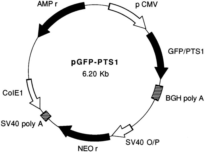
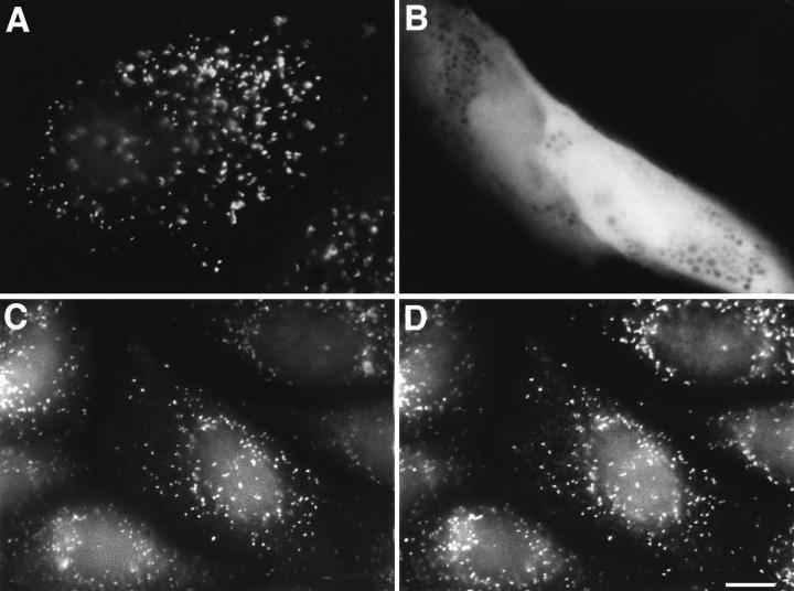
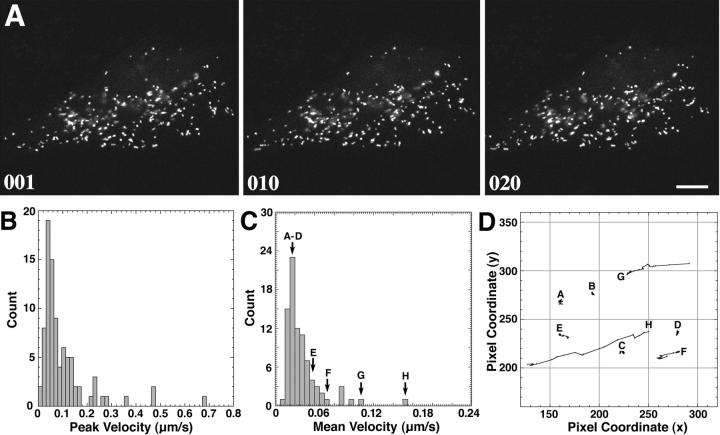
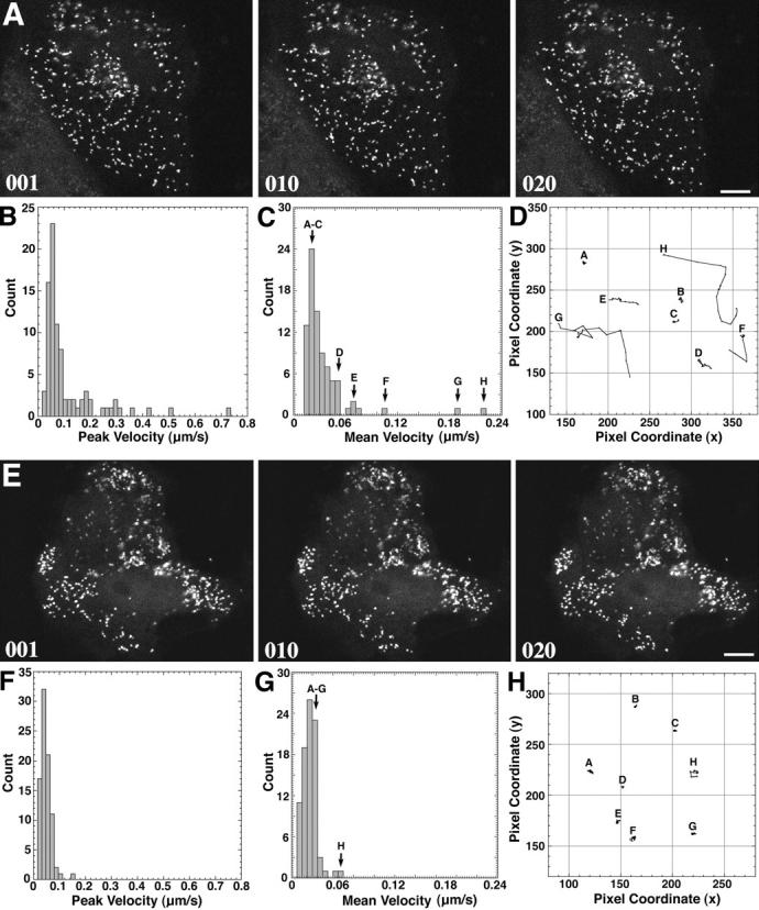
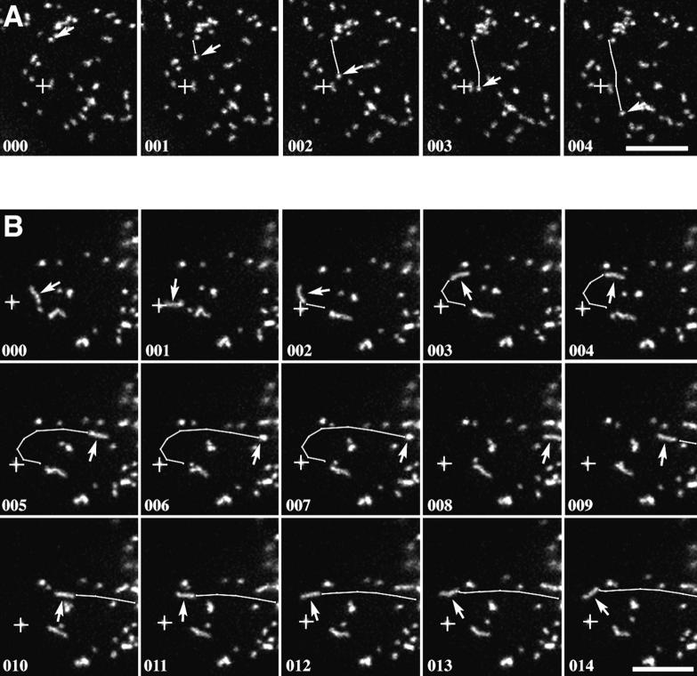
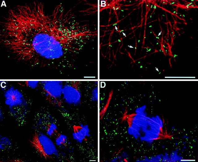
Peroxisomes in living CV1 cells were visualized by targeting the green fluorescent protein (GFP) to this subcellular compartment through the addition of a COOH-terminal peroxisomal targeting signal 1 (GFP-PTS1). The organelle dynamics were examined and analyzed using time-lapse confocal laser scanning microscopy. Two types of movement could be distinguished: a relatively slow, random, vibration-like movement displayed by the majority (approximately 95%) of the peroxisomes, and a saltatory, fast directional movement displayed by a small subset (approximately 5%) of the peroxisomes. In the latter instance, peak velocities up to 0.75 micron/s and sustained directional velocities up to 0.45 micron/s over 11.5 microns were recorded. Only the directional type of motion appeared to be energy dependent, whereas the vibrational movement continued even after the cells were depleted of energy. Treatment of cells, transiently expressing GFP-PTS1, with microtubule-destabilizing agents such as nocodazole, vinblastine, and demecolcine clearly altered peroxisome morphology and subcellular distribution and blocked the directional movement. In contrast, the microtubule-stabilizing compound paclitaxel, or the microfilament-destabilizing drugs cytochalasin B or D, did not exert these effects. High resolution confocal analysis of cells expressing GFP-PTS1 and stained with anti-tubulin antibodies revealed that many peroxisomes were associated with microtubules. The GFP-PTS1-labeled peroxisomes were found to distribute themselves in a stochastic, rather than ordered, manner to daughter cells at the time of mitosis.
Figures
Similar articles
-
Interaction of microtubules with peroxisomes. Tubular and spherical peroxisomes in HepG2 cells and their alterations induced by microtubule-active drugs.Eur J Cell Biol. 1996 Jan;69(1):24-35. Eur J Cell Biol. 1996. PMID: 8825021
-
Visualization of peroxisomes in living plant cells reveals acto-myosin-dependent cytoplasmic streaming and peroxisome budding.Plant Cell Physiol. 2002 Apr;43(4):384-92. doi: 10.1093/pcp/pcf045. Plant Cell Physiol. 2002. PMID: 11978866
-
Distribution and characterization of peroxisomes in Arabidopsis by visualization with GFP: dynamic morphology and actin-dependent movement.Plant Cell Physiol. 2002 Mar;43(3):331-41. doi: 10.1093/pcp/pcf037. Plant Cell Physiol. 2002. PMID: 11917088
-
Simultaneous visualization of peroxisomes and cytoskeletal elements reveals actin and not microtubule-based peroxisome motility in plants.Plant Physiol. 2002 Mar;128(3):1031-45. doi: 10.1104/pp.011018. Plant Physiol. 2002. PMID: 11891258 Free PMC article.
-
Targeted fluorescent probes in peroxisome function.Histochem J. 2001 Feb;33(2):65-9. doi: 10.1023/a:1017927728892. Histochem J. 2001. PMID: 11432641 Review.
Cited by
-
Peroxisome distribution along the crypt-villus axis of the guinea pig small intestine.Mol Cell Biochem. 2000 Jan;203(1-2):119-26. doi: 10.1023/a:1007052003143. Mol Cell Biochem. 2000. PMID: 10724340
-
Enzyme-Instructed Assemblies Enable Mitochondria Localization of Histone H2B in Cancer Cells.Angew Chem Int Ed Engl. 2020 Jun 8;59(24):9330-9334. doi: 10.1002/anie.202000983. Epub 2020 Mar 31. Angew Chem Int Ed Engl. 2020. PMID: 32119754 Free PMC article.
-
Live-cell analysis of a green fluorescent protein-tagged herpes simplex virus infection.J Virol. 1999 May;73(5):4110-9. doi: 10.1128/JVI.73.5.4110-4119.1999. J Virol. 1999. PMID: 10196307 Free PMC article.
-
Guidelines for the use and interpretation of assays for monitoring autophagy (4th edition)1.Autophagy. 2021 Jan;17(1):1-382. doi: 10.1080/15548627.2020.1797280. Epub 2021 Feb 8. Autophagy. 2021. PMID: 33634751 Free PMC article.
-
Bidirectional translocation of neurofilaments along microtubules mediated in part by dynein/dynactin.Mol Biol Cell. 2000 Oct;11(10):3495-508. doi: 10.1091/mbc.11.10.3495. Mol Biol Cell. 2000. PMID: 11029051 Free PMC article.
References
-
- Allan V. Organelle movement. Dynactin: portrait of a dynein regulator. Curr Biol. 1994;4:1000–1002. - PubMed
-
- Baumann O, Murphy DB. Microtubule-associated movement of mitochondria and small particles in Acanthamoeba castellanii. . Cell Motil Cytoskeleton. 1995;32:305–317. - PubMed
-
- Birky CW. The partitioning of cytoplasmic organelles at cell division. Int Rev Cytol. 1983;15:49–89. - PubMed
-
- Cole NB, Lippincott-Schwartz J. Organization of organelles and membrane traffic by microtubules. Curr Opin Cell Biol. 1995;7:55–64. - PubMed
-
- Cubitt AB, Heim R, Adams SR, Boyd AE, Gross LA, Tsien RY. Understanding, improving and using green fluorescent proteins. Trends Biochem Sci. 1995;20:448–455. - PubMed
Publication types
MeSH terms
Substances
Grants and funding
LinkOut - more resources
Full Text Sources
Other Literature Sources
