. 1997 Jan 13;136(1):177-91.
doi: 10.1083/jcb.136.1.177.

Phosphoregulation of an inner dynein arm complex in Chlamydomonas reinhardtii is altered in phototactic mutant strains

Affiliations

Phosphoregulation of an inner dynein arm complex in Chlamydomonas reinhardtii is altered in phototactic mutant strains

S J King et al. J Cell Biol. .

Abstract

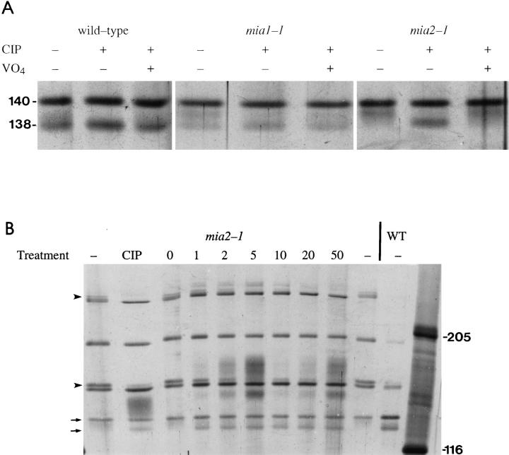
To gain a further understanding of axonemal dynein regulation, mutant strains of Chlamydomonas reinhardtii that had defects in both phototactic behavior and flagellar motility were identified and characterized. ptm1, ptm2, and ptm3 mutant strains exhibited motility phenotypes that resembled those of known inner dynein arm region mutant strains, but did not have biochemical or genetic phenotypes characteristic of other inner dynein arm mutations. Three other mutant strains had defects in the f class of inner dynein arms. Dynein extracts from the pf9-4 strain were missing the entire f complex. Strains with mutations in pf9/ida1, ida2, or ida3 failed to assemble the f dynein complex and did not exhibit phototactic behavior. Fractionated dynein from mia1-1 and mia2-1 axonemes exhibited a novel f class inner dynein arm biochemical phenotype; the 138-kD f intermediate chain was present in altered phosphorylation forms. In vitro axonemal dynein activity was reduced by the mia1-1 and mia2-1 mutations. The addition of kinase inhibitor restored axonemal dynein activity concomitant with the dephosphorylation of the 138-kD f intermediate chain. Dynein extracts from uni1-1 axonemes, which specifically assemble only one of the two flagella, contained relatively high levels of the altered phosphorylation forms of the 138-kD intermediate chain. We suggest that the f dynein complex may be phosphoregulated asymmetrically between the two flagella to achieve phototactic turning.


Figures

Figure 1
Figure 1
Scanned image of a genomic Southern blot that contains DNA from three mutant strains. The blot was probed with a radiolabeled pARG7.8 vector fragment. SalI restriction endonuclease–digested AT2-121 (A) and AT1-25 (B) genomes contained a single insertion, whereas the AT1-2 (C) genome contained two insertions. (Left) Positions of size standards in kb.
Figure 2
Figure 2
Silver-stained SDSPAGE gels of the HPLCfractionated dynein heavy and intermediate chains from wild-type (A) and pf9-4 (B) axonemes. The HPLC fractions are labeled for each lane. hse, high salt extract; ax, whole axonemes. Several dynein peaks contained polypeptides from neighboring dynein peaks. The polypeptides that were part of the particular complex separated in each peak are labeled with a dot on the right of the band. The F fraction of pf9-4 dynein extracts was missing two heavy and three intermediate f dynein polypeptides; the expected positions of these bands are shown by asterisks. This gel did not resolve the 110-kD f intermediate chain, but it was missing in other gels (data not shown). The positions of 205- and 116-kD standards are indicated.
Figure 3
Figure 3
Silver-stained gel showing HPLC fractionated polypeptides from wild-type, mia1-1, and mia2-1 dynein extracts. One fraction of wild-type and three consecutive fractions of mia1-1 and mia2-1 extracts are shown. Polypeptides of the αβ outer arm and g inner arm dyneins cofractionated to some extent with f dynein (Fig. 2; Kagami and Kamiya, 1992; Gardner et al., 1994). The wild-type fraction was underloaded compared with the other fractions. When wild-type fractions were overloaded, no additional bands were observed as in mia fractions. (Arrows, left) Positions of the 140-kD and 138-kD intermediate chains of the wild-type f dynein complex. (Arrowhead, right) Position(s) of the new polypeptide species seen in mia1-1 and mia2-1 dynein extracts. The positions of 205- and 116-kD standards are indicated.
Figure 4
Figure 4
Phosphatase treatment alters the electrophoretic mobility of f dynein polypeptides. (A) Silverstained gels of the f dynein polypeptides from wild-type, mia1-1, and mia2-1 dynein extracts after treatment with CIP. Only the region of the gel that contained the f dynein 140- and 138-kD intermediate polypeptides is shown. The wild-type lanes were overloaded to show the faint smear of polypeptides that extended upward from the 138-kD polypeptide. Positions of the 140- and 138kD polypeptides (left). The presence (+) or absence (−) of phosphatase (CIP) and 5 μM sodium orthovanadate (VO4) is indicated. (B) Silver-stained gel showing the electrophoretic mobility of F fraction polypeptides. The dynein heavy chains are not shown. The far right lane contains 205- and 116-kD standards. The next to last lane on the right contains wild-type F fraction polypeptides. The remaining lanes contain equal amounts of mia2-1 F fraction polypeptides. Phosphatase addition is indicated above each lane; −, no treatment; CIP, calf intestinal phosphatase treatment; calcineurin treatment, numbers correspond to units of calcineurin added. (Arrows) Positions of the 140- and 138-kD polypeptides. The migration of two additional non-f dynein polypeptides was affected by calcineurin treatment; these bands are indicated by arrowheads.
Figure 5
Figure 5
The 74-kD region of silver-stained two-dimensional gels. (Arrow) Position of the 74-kD polypeptide in wild-type axonemes (A); (arrowhead) expected position in mia1-1 axonemes (B). All twodimensional gels are shown with acidic pIs to the right. Other polypeptide variability seen in the two-dimensional gels of Figs. 5–7 was not consistent in other gels and in other axonemal preparations.
Figure 6
Figure 6
The 35-kD region of silver-stained two-dimensional gels of axonemes from different strains. (A) wild-type; (B) pf9-2; (C) mia1-1; (D) mia2-1; (E) wild-type and mia2-1; (F) mia2-1R-14. (A, arrows) Positions of the 35- and 34-kD polypeptides discussed in the text. (open arrowheads, pointing down) Expected position of the 35-kD polypeptide. (open arrowheads, pointing up) Expected position of the 34-kD polypeptide. (D, E, and F, small arrows) Positions of the novel polypeptide found in mia2-1 axonemes. (F, black arrowhead) Return of the 34-kD polypeptide in mia2-1R-14 axonemes.
Figure 7
Figure 7
The 138-kD region of silver-stained two-dimensional gels of axonemes from different strains. (A) wild-type; (B) mia11; (C and D) mia1-1 spf1-1; (E) pf9-2; (F) mia2-1; (G and H) mia2-1 spf1-1; (I) mia2-1R-14. The axonemes in D and H were treated with HA-1004. (Arrows) Positions of the 138- and 110-kD f polypeptides in wildtype axonemes (A). The series of 138-kD polypeptide spots is found just below a more prominent axonemal polypeptide series. (Filled arrowheads) Position of the most basic form of the 138kD polypeptide found in the corresponding axonemes. (Open arrowheads) Expected positions of the 138- and 110-kD polypeptides in pf9-2 axonemes (E).
Figure 8
Figure 8
Sliding velocities of axonemes from various strains. The sliding velocities (in μm/s ± SD) of axonemes without (top values; labeled control) and with HA-1004 (bottom values; labeled ±KI) treatment are shown in bold type. The value by each bracket is the P value of an unpaired two-tailed t test between the corresponding sliding velocity distributions. P values of <0.05 were considered significant. 27–31 independent events were measured for each condition.
Figure 9
Figure 9
Silver-stained gel showing HPLC-fractionated polypeptides from uni1-1 dynein extracts. Two consecutive fractions are shown. (Arrowhead) Positions of the new polypeptide species seen in the dynein extract. The 138-kD polypeptide phenotype in uni1-1 dynein extracts is similar to that seen in mia1-1 dynein extracts (Figs. 3 and 4). The positions of 205- and 116-kD standards are indicated.
Figure 10
Figure 10
Schematic model of asymmetric phosphoregulation of f dynein activity and its role in phototaxis. When the eyespot is lighted (A), the rhodopsin signal is transduced to the flagellar membrane where Ca++ ion channels would be opened (1). Elevated Ca++ concentrations would increase the activity of the phosphatase relative to the kinase in the trans flagellum (2). This would result in the dephosphorylation of the 138-kD intermediate chain and subsequent activation of the f dynein complex. The trans flagellum would then beat with an increased front amplitude (shaded area under flagella) relative to the cis flagellum, and the cell would turn toward the light source (3). As the cell rotates, the eyespot becomes shaded (B) and the flagellar Ca++ ion channels would be closed (1). The resultant reduction in Ca++ ion concentration would increase the activity of the kinase relative to the phosphatase (2). This would lead to phosphorylation of the 138-kD intermediate chain and the deactivation of the f dynein complex. The trans flagellum would then beat with a decreased front amplitude relative to the cis flagellum, and the cell would continue to turn toward the light source (3).

Similar articles

Cited by

References

    1. Adams GMW, Huang B, Piperno G, Luck DJL. Central-pair microtubular complex of Chlamydomonasflagella: polypeptide composition as revealed by analysis of mutants. J Cell Biol. 1981;91:69–76. - PMC - PubMed
    1. Barkalow K, Hamasaki T, Satir P. Regulation of 22S dynein by a 29-kD light chain. J Cell Biol. 1994;126:727–735. - PMC - PubMed
    1. Blum H, Beier H, Gross HJ. Improved silver staining of plant proteins, RNA and DNA in polyacrylamide gels. Electrophoresis. 1987;8:93–97.
    1. Brokaw CJ, Kamiya R. Bending patterns of Chlamydomonasflagella: IV. Mutants with defects in inner and outer dynein arms indicate differences in dynein arm function. Cell Motil. 1987;8:68–75. - PubMed
    1. Cohen P. The role of protein phosphorylation in neural and hormonal control of cellular activity. Nature (Lond) 1982;296:613–620. - PubMed

Publication types