. 1997 Jan 13;136(1):205-13.
doi: 10.1083/jcb.136.1.205.

Bcl-2 lies downstream of parathyroid hormone-related peptide in a signaling pathway that regulates chondrocyte maturation during skeletal development

Affiliations

Bcl-2 lies downstream of parathyroid hormone-related peptide in a signaling pathway that regulates chondrocyte maturation during skeletal development

M Amling et al. J Cell Biol. .

Abstract

Parathyroid hormone-related peptide (PTHrP) appears to play a major role in skeletal development. Targeted disruption of the PTHrP gene in mice causes skeletal dysplasia with accelerated chondrocyte maturation (Amizuka, N., H. Warshawsky, J.E. Henderson, D. Goltzman, and A.C. Karaplis. 1994. J. Cell Biol. 126:1611-1623; Karaplis, A.C., A. Luz, J. Glowacki, R.T. Bronson, V.L.J. Tybulewicz, H.M. Kronenberg, and R.C. Mulligan. 1994. Genes Dev. 8: 277-289). A constitutively active mutant PTH/PTHrP receptor has been found in Jansen-type human metaphyseal chondrodysplasia, a disease characterized by delayed skeletal maturation (Schipani, E., K. Kruse, and H. Jüppner. 1995. Science (Wash. DC). 268:98-100). The molecular mechanisms by which PTHrP affects this developmental program remain, however, poorly understood. We report here that PTHrP increases the expression of Bcl-2, a protein that controls programmed cell death in several cell types, in growth plate chondrocytes both in vitro and in vivo, leading to delays in their maturation towards hypertrophy and apoptotic cell death. Consequently, overexpression of PTHrP under the control of the collagen II promoter in transgenic mice resulted in marked delays in skeletal development. As anticipated from these results, deletion of the gene encoding Bcl-2 leads to accelerated maturation of chondrocytes and shortening of long bones. Thus, Bcl-2 lies downstream of PTHrP in a pathway that controls chondrocyte maturation and skeletal development.


Figures

Figure 1
Figure 1
Expression of Bcl-2 and Bax in endochondral bone formation. (A) The normal growth plate shows a typical zonal structure of differentiating chondrocytes. Proliferating chondrocytes (P) progressively enlarge to prehypertrophic chondrocytes (PH), and further differentiate into hypertrophic chondrocytes (H). (B) Age-matched growthplates were labeled by immunofluorescence for collagen type X, which serves as a specific marker in the chondrocyte differentiation process. Intracellular expression of collagen X is characteristic of prehypertrophic chondrocytes, whereas the dense, pericellular matrix labeling shown in red is found solely in the hypertrophic zone. (C) Highest levels of Bcl-2 protein are detected in late proliferative and prehypertrophic chondrocytes, whereas, (D), hypertrophic chondrocytes exhibit an increased Bax protein expression. Age-matched frozen sections were labeled for DNA fragmentation by TUNEL. (E) Nick end labeling of DNA fragments was observed only in fully differentiated hypertrophic chondrocytes, at the level where resorption of cartilage and replacement by bone is taking place.
Figure 2
Figure 2
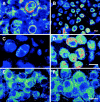
PTHrP increases Bcl-2 expression in chondrocytes in vitro. (A) Murine chondrocytes, cultured for 24 d as high-density monolayers, differentiated in vitro into hypertrophic chondrocytes with the typical extracellular matrix pattern of collagen X (compare to Fig. 1 B). (B) In contrast, treatment of these cultures with PTHrP 1-37 resulted in an inhibition of terminal differentiation and an accumulation of prehypertrophic chondrocytes, as confirmed by the pathognomonic intracellular collagen X expression. After 12 d in culture, a time at which treated and untreated cultures show no apparent morphological differences, (C) the untreated chondrocytes show only low cytoplasmic Bcl-2 protein expression, whereas, (D), a marked increase in Bcl-2 expression is visible after PTHrP treatment. In contrast, the same treatment had no detectable effect on the level of Bax protein expression (E, control; F, treated). Bars, 10 μm.
Figure 3
Figure 3
PTHrP effect on Bcl-2 is restricted to chondrocytes. Western blotting analysis revealed that treatment of murine chondrocytes in vitro (cultured for 12 d) with PTHrP 1-37 resulted in a marked increase in Bcl-2 expression. In contrast, the same treatment had no detectable effect on Bcl-2 levels in primary osteoblasts, obtained from 3-d–old mouse calvaria, and a kidney cell line (CAKI), both of which express the PTH/PTHrP receptor. The blots were then stripped of antibody and reprobed with anti-actin antibody to confirm that equal amounts of protein were loaded in each corresponding treated and untreated lane.
Figure 4
Figure 4
Targeted overexpression of PTHrP to chondrocytes in transgenic animals leads to increased Bcl-2 expression and delayed chondrocyte maturation. (A) Metatarsals of a normal 6-d–old mouse, showing the zonal structures of a growth plate, trabecular and cortical bone, and the presence of a marrow cavity. In contrast (B), the metatarsal of the col II–PTHrP transgenic littermate demonstrates a delay in chondrocyte differentiation, an accumulation of prehypertrophic chondrocytes (as confirmed by intracellular collagen X expression, data not shown), and the complete absence of bone formation. At a higher magnification of the insets in A and B, the progression of chondrocyte differentiation is visible in the normal growth plate: proliferating cells differentiate first into prehypertrophic cells, and after further enlargement, they finally die at the border to bone formation (compare to Fig. 1) (C). In contrast, the same region in the metatarsal of the col II–PTHrP transgenic mouse shows that the cells accumulate at the prehypertrophic stage (D). Frozen sections of the corresponding growth plate regions labeled for Bcl-2 and Bax by immunofluorescence demonstrate the normal pattern of Bcl-2 expression in the growth plate, with highest levels in the zone of prehypertrophic chondrocytes, (E). In contrast, in the col II–PTHrP transgenic animals, not only the number of chondrocytes expressing Bcl-2 but also the level of Bcl-2 expression are markedly increased (F); panels G and H demonstrate that Bax expression is not affected in the col II–transgenic animals (H) and is comparable to normal levels in prehypertrophic chondrocytes (G).
Figure 5
Figure 5
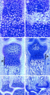
Bcl-2 directly affects skeletal development and endochondral bone formation. bcl-2 knockout mice exhibit accelerated chondrocyte differentiation. (A) While under normal conditions at day 1, the foot middle phalanx is still a cartilaginous model. (B) In bcl-2 knockout littermates, vascular invasion, progressive replacement of cartilage by bone, and homing of bone marrow cells has already taken place. (D) This acceleration of chondrocyte differentiation leads to a reduction in the growth plate thickness, mostly in the proliferative zone (P), shown here for the distal metatarsal bone of a 6-d–old bcl-2 knockout mouse as compared to a 6-d–old normal control (C). Even more striking is the almost complete loss of cartilage at the proximal end of the metatarsal bones (day 6) in the bcl-2 −/− (F), while in the normal mice, the present zone of chondrocytes is still a resource of bone formation (E). Consequently, bcl-2 knockout mice are markedly smaller than control littermates (G) (contact x-ray of 60-d-old mice; on the right bcl-2 −/−, on the left normal control littermate), and the absence of Bcl-2 leads to a significant decrease (15%–20%) in overall bone length (H). Significant levels of P < 0.05, Student's t test, are indicated by asterisks.
Figure 5
Figure 5
Bcl-2 directly affects skeletal development and endochondral bone formation. bcl-2 knockout mice exhibit accelerated chondrocyte differentiation. (A) While under normal conditions at day 1, the foot middle phalanx is still a cartilaginous model. (B) In bcl-2 knockout littermates, vascular invasion, progressive replacement of cartilage by bone, and homing of bone marrow cells has already taken place. (D) This acceleration of chondrocyte differentiation leads to a reduction in the growth plate thickness, mostly in the proliferative zone (P), shown here for the distal metatarsal bone of a 6-d–old bcl-2 knockout mouse as compared to a 6-d–old normal control (C). Even more striking is the almost complete loss of cartilage at the proximal end of the metatarsal bones (day 6) in the bcl-2 −/− (F), while in the normal mice, the present zone of chondrocytes is still a resource of bone formation (E). Consequently, bcl-2 knockout mice are markedly smaller than control littermates (G) (contact x-ray of 60-d-old mice; on the right bcl-2 −/−, on the left normal control littermate), and the absence of Bcl-2 leads to a significant decrease (15%–20%) in overall bone length (H). Significant levels of P < 0.05, Student's t test, are indicated by asterisks.

References

    1. Arends MJ, Morris RG, Wyllie AH. Apoptosis. The role of the endonuclease. Am J Pathol. 1990;136:593–608. - PMC - PubMed
    1. Allsopp TE, Wyatt S, Paterson HF, Davies AM. The proto- oncogene bcl-2can selectively rescue neurotrophic factor–dependent neurons from apoptosis. Cell. 1993;73:295–307. - PubMed
    1. Amizuka N, Warshawsky H, Henderson JE, Goltzman D, Karaplis AC. Parathyroid hormone–related, peptide-depleted mice show abnormal epiphyseal cartilage development and altered endochondral bone formation. J Cell Biol. 1994;126:1611–1623. - PMC - PubMed
    1. Blanco FJ, Ochs RL, Schwarz H, Lotz M. Chondrocyte apoptosis induced by nitric oxide. Am J Pathol. 1995;146:75–85. - PMC - PubMed
    1. Boise LH, González-García M, Postema CE, Ding L, Lindsten T, Turca LA, Mao X, Nunez G, Thompson CB. bcl-x, a bcl-2–related gene that functions as a dominant regulator of apoptotic cell death. Cell. 1993;74:597–608. - PubMed

Publication types

MeSH terms