Review
. 1997 Jan 21;94(2):377-84.
doi: 10.1073/pnas.94.2.377.

The dynamins: redundant or distinct functions for an expanding family of related GTPases?

Affiliations
Review

The dynamins: redundant or distinct functions for an expanding family of related GTPases?

R Urrutia et al. Proc Natl Acad Sci U S A. .

Abstract

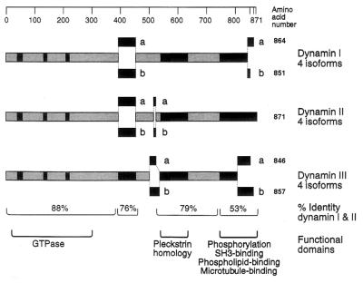
In the 7 years since dynamin was first isolated from bovine brain in search of novel microtubule-based motors, our understanding of this enzyme has expanded significantly. We now know that brain dynamin belongs to a family of large GTPases, which mediate vesicle trafficking. Furthermore, this enzymatic activity is markedly increased through association with microtubules, acidic phospholipids, and certain regulatory proteins that contain Src homology 3 (SH3) domains. From functional, genetic, and cellular manipulations, it is now generally accepted that dynamin participates in the endocytic uptake of receptors, associated ligands, and plasma membrane following an exocytic event. These observations have confirmed at least one function of dynamin that was predicted from seminal studies on a pleiotropic mutant, shibire(ts) (shi(ts)) in Drosophila melanogaster. Of equal interest is the finding that there are multiple dynamin gene products, including two that are expressed in a tissue-specific manner, and they share marked homology with a larger family of distinct but related proteins. Therefore, it is attractive to speculate that the different dynamins may participate in related cellular functions, such as distinct endocytic processes and even secretion. In turn, dynamin could play an important role in cell growth, cell spreading, and neurite outgrowth. The purpose of this review is to enumerate on the expansive dynamin literature and to discuss the nomenclature, expression, and putative functions of this growing and interesting family of proteins.


Figures

Figure 1
Figure 1
Endocytosis in neurons and epithelial cells is structurally altered in the Drosophila mutant shits. (a and b) Electron micrographs of a typical coxal synapse of a shits fly at 19°C (permissive temperature), which is characterized by numerous SVs, mitochondria (M), and a presynaptic dense body (DB). (b) A similar synapse at the restrictive temperature of 29°C. Note the loss of SVs and the appearance of several small invaginations of the plasma membrane, which lack clathrin but possess an electron-dense ring or collar around their necks (arrowheads). (c) A higher magnification of a synapse containing collared pits at 29°C. (d) In contrast to neuronal cells, long labyrinthine channels are formed in the cortical region of a shits garland cell at the restrictive temperature that lack collars. Cells have been impregnated with tannic acid to show that these structures (arrows) are continuous with the plasma membrane and end with a clathrin-coated pit (arrowheads). Few discrete coated vesicles are seen. [ac were reproduced with permission from Koenig and Ikeda (4) (Copyright 1989, Soc. Neurosci.); and d was reproduced with permission from Kosaka and Ikeda (5) (Copyright 1983, Rockefeller Univ. Press).]
Figure 2
Figure 2
Domain structure of the dynamin family. The products of three distinct mammalian genes have been characterized and give rise to multiple proteins that are generated by alternative splicing of the mRNA. DynI is expressed in neuronal tissue, DynII is expressed in all tissues, and DynIII is expressed in the brain, lung, and testis. Dynamin isoform nomenclature makes use of the alternative splice sites; thus the 96-kDa form of DynI is either DynIaa or DynIba, while the 94-kDa form is DynIab or DynIbb. [Reproduced with permission from Robinson et al. (19) (Copyright 1994, Elsevier Science).]
Figure 3
Figure 3
Manipulation of mammalian cells to induce endocytic alterations reminiscent of the shits phenotype. (a) Transmission electron microscopy of transfected HeLa cells overexpressing a mutant DynI protein. In these cells, long membranous invaginations which are continuous with the plasma membrane can be seen (arrows). (b and c) Transmission electron microscopy of isolated rat brain synaptosomes incubated with GTP[γS]. Under these conditions, numerous membrane invaginations can be observed extending from the synaptosomal membrane. Numerous dense staining striations similar to the collars observed in shits can be seen encircling the neck of each invagination (arrowheads). These striations are labeled extensively with a monoclonal antibody to DynI (d). Numerous immunogold particles can be seen around the shaft of each tubule (arrowheads). [a was reproduced with permission from Damke et al. (12) (Copyright 1994, Rockefeller Univ. Press); and bd were reproduced with permission from Takei et al. (20) (Copyright 1995, Macmillan Magazines).]
Figure 4
Figure 4
DynI assembles into stacked ring structures in the absence or presence of Mts. (a) Negative-stain electron microscopy of individual disassembled dynamin oligomers in a low-salt buffer. With an increase in dynamin protein concentration (b), the oligomers self-associate to form elongated stacks of rings. Similar stacked-ring arrangements are seen when purified taxol-stabilizing Mts (c) are incubated with purified brain dynamin and viewed by quick-freeze deep-etch electron microscopy (d). Dynamin can also cross-link Mts as shown by negatively stained preparations of taxol-stabilized Mts with purified brain dynamin. Dynamin molecules can be seen as periodic striations connecting adjacent Mts into bundles (e and f). [a and b were reproduced with permission from Hinshaw and Schmid (33) (Copyright 1995, Macmillan Magazines); c and d were reproduced with permission from Maeda et al. (31) (Copyright 1992, Am. Soc. Cell Biol.); and e and f were reproduced with permission from Shpetner and Vallee (34) (Copyright 1989, Cell Press).]
Figure 5
Figure 5
Cellular localizations of the dynamin superfamily by immunofluorescence microscopy. (a) Cultured HeLa cells stained with a monoclonal antibody to dynamin. Numerous punctate structures (arrows) are seen throughout the cytoplasm as well as a prominent juxtanuclear labeling (arrowheads). (b) Immunostaining of cultured human fibroblasts stained with an affinity-purified polyclonal antibody made to a region conserved among the dynamins. An extensive reticular Golgi network can be seen surrounding the nucleus. (c) Cultured human fibroblasts stained with a polyclonal antibody (gift of T. Stevens, University of Oregon) to the yeast GTP-binding protein Vps1p. Similar to the staining pattern seen with the dynamin antibodies, elaborate reticular Golgi structures (arrows) are labeled in each cell. (d and e) Immunolocalization of dynamin in sea urchin eggs using an antibody made to a dynamin-related protein purified from these eggs. (d) Dynamin localization in the cortex of these eggs appears as diffuse and cytoplasmic in unfertilized eggs. (e) Immediately after fertilization, the honeycomb cortical granule staining pattern is disrupted, and two populations of brightly staining spots begin to appear throughout the periphery, suggesting an association with cortical membrane. (f) Immunostaining of cultured rat hippocampal neurons with affinity-purified antibodies to conserved domains of dynamin show bright punctate staining at specific locations along extended neurites (arrows). (g and h) Higher magnification images of growth cones from these cells show a restricted, yet prominent, dynamin localization at the very tip of these processes (arrows). [a was reproduced with permission from Damke et al. (12) (Copyright 1994, Rockefeller Univ. Press); b and c were reproduced with permission from Henley and McNiven (61) (Copyright 1996, Rockefeller Univ. Press); d and e were reproduced with permission from Faire and Bonder (36) (Copyright 1993, Academic Press); and f–h were reproduced with permission from E. Torre and M.A.M. (unpublished observations).

References

    1. Grigliatti T A, Hall L, Rosenbluth R, Suzuki D T. Mol Gen Genet. 1973;120:107–114. - PubMed
    1. Poodry C A. Dev Biol. 1990;138:464–472. - PubMed
    1. Kosaka T, Ikeda K. J Neurobiol. 1983;14:207–225. - PubMed
    1. Koenig J H, Ikeda K. J Neurosci. 1989;9:3844–3860. - PMC - PubMed
    1. Kosaka T, Ikeda K. J Cell Biol. 1983;97:499–507. - PMC - PubMed

Publication types

LinkOut - more resources