. 1997 Jan 21;94(2):479-84.
doi: 10.1073/pnas.94.2.479.

The thioredoxin binding domain of bacteriophage T7 DNA polymerase confers processivity on Escherichia coli DNA polymerase I

Affiliations

The thioredoxin binding domain of bacteriophage T7 DNA polymerase confers processivity on Escherichia coli DNA polymerase I

E Bedford et al. Proc Natl Acad Sci U S A. .

Abstract

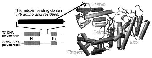
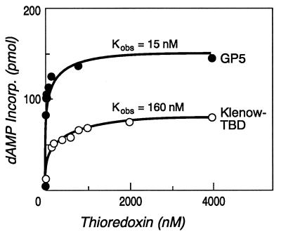
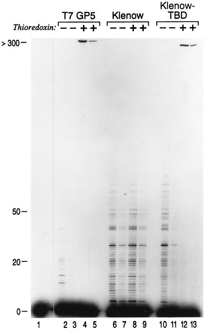
Bacteriophage T7 DNA polymerase shares extensive sequence homology with Escherichia coli DNA polymerase I. However, in vivo, E. coli DNA polymerase I is involved primarily in the repair of DNA whereas T7 DNA polymerase is responsible for the replication of the viral genome. In accord with these roles, T7 DNA polymerase is highly processive while E. coli DNA polymerase I has low processivity. The high processivity of T7 DNA polymerase is achieved through tight binding to its processivity factor, E. coli thioredoxin. We have identified a unique 76-residue domain in T7 DNA polymerase responsible for this interaction. Insertion of this domain into the homologous site in E. coli DNA polymerase I results in a dramatic increase in the processivity of the chimeric DNA polymerase, a phenomenon that is dependent upon its binding to thioredoxin.


Figures

Figure 1
Figure 1
Location of the thioredoxin binding domain (TBD) in T7 and Klenow–TBD DNA polymerases. (Right) The x-ray crystal structure of Klenow DNA polymerase is shown (10), modeled using the program setor (11). (Left) The alignment of the H–H1 region in T7 DNA polymerase and E. coli DNA polymerase I is shown (7). The unique 76-residue region in T7 DNA polymerase, located between helices H and H1, is indicated by the dark rectangle. In the chimeric Klenow–TBD DNA polymerase, this domain has been inserted at the tip of the thumb with the deletion of seven residues from E. coli DNA polymerase, as indicated by the arrow.
Figure 2
Figure 2
Stimulation of DNA synthesis by thioredoxin. The amount of DNA synthesis in the presence of increasing concentrations of thioredoxin was determined in reactions catalyzed by either T7 DNA polymerase (GP5) or Klenow–TBD DNA polymerase (Klenow–TBD). The amount of [32P]dAMP incorporated into primed single-stranded M13 mGP1-2 DNA was determined as described. The molar ratio of DNA polymerase to primer–template was 1:10 for all reactions.
Figure 3
Figure 3
Surface plasmon resonance analysis of Klenow–TBD polymerase/thioredoxin interaction. (A) The binding of thioredoxin to a sensor chip that has antithioredoxin mAbs covalently attached to its surface. The open arrow indicates the start of the injection of thioredoxin, and the filled arrow indicates the start of the buffer wash. The number of RU that remain after the initial sharp decay at the start of the buffer wash are indicated by the discontinuous arrows. (B) The binding of Klenow DNA polymerase to a chip containing thioredoxin bound to the mAbs. (C) The binding of Klenow–TBD DNA polymerase to a chip containing thioredoxin bound to the mAbs.
Figure 4
Figure 4
The effect of thioredoxin on the processivity of Klenow–TBD polymerase. DNA polymerase reactions were carried out using a 5′ 32P-labeled primer annealed to M13 mGP1-2 DNA in the presence and absence of thioredoxin. Lane 1 contains the starting primer–template in the absence of any DNA polymerase. In the even-numbered lanes (bottom) the primer–template was present at a 50-fold molar excess over the DNA polymerase, and in the odd-numbered lanes the primer–template present at a 250-fold molar excess over the DNA polymerase. In lanes 2–5 the reactions were carried out using T7 DNA polymerase (T7 GP5). In lanes 6–9 the reactions were carried out using Klenow DNA polymerase (Klenow), and in lanes 10–13 the reactions were carried out using Klenow–TBD DNA polymerase (Klenow–TBD). The products were separated on a denaturing 8% polyacrylamide gel. After electrophoresis, the gel was dried and autoradiographed. The mobility of primers that have been extended by the indicated numbers of nucleotides is shown on the left.

References

    1. Kornberg A, Baker T A. DNA Replication. 2nd Ed. New York: Freeman; 1992.
    1. Tabor S, Huber H E, Richardson C C. J Biol Chem. 1987;262:16212–16223. - PubMed
    1. Modrich P, Richardson C C. J Biol Chem. 1975;250:5515–5522. - PubMed
    1. Mark D, Richardson C C. Proc Natl Acad Sci USA. 1976;73:780–784. - PMC - PubMed
    1. Huber H E, Russel M, Model P, Richardson C C. J Biol Chem. 1986;261:15006–15012. - PubMed

Publication types

LinkOut - more resources