. 1997 Jan 21;94(2):485-90.
doi: 10.1073/pnas.94.2.485.

Probing the environment along the protein import pathways in yeast mitochondria by site-specific photocrosslinking

Affiliations

Probing the environment along the protein import pathways in yeast mitochondria by site-specific photocrosslinking

T Kanamori et al. Proc Natl Acad Sci U S A. .

Abstract

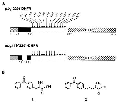
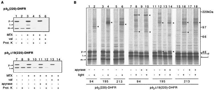
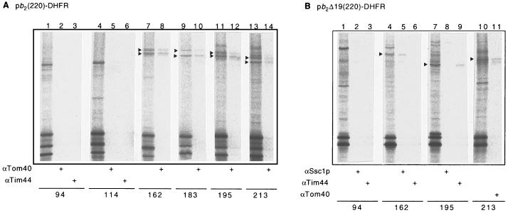
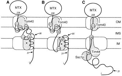
Artificially aminoacylated suppressor tRNAs were used to introduce photoreactive amino acids into model mitochondrial precursor proteins to probe the environment along the protein import pathway. Amino acids with benzophenone side chains of various lengths [DL-2-amino-3-(p-benzoylphenyl)propanoic acid (1) and DL-2-amino-5-(p-benzoylphenyl)pentanoic acid (2)] were incorporated at specific sites throughout the cytochrome b2-dihydrofolate reductase fusion proteins, pb2(220)-DHFR and pb2 delta 19(220)-DHFR, which were destined for the intermembrane space and the matrix in mitochondria, respectively. In vitro import of pb2(220)-DHFR and pb2 delta 19(220)-DHFR bearing 1 or 2 into isolated yeast mitochondria was arrested so that the N terminus reached the intermembrane space or the matrix, respectively, while the DHFR domain remained at the mitochondrial surface. The matrix-targeted pb2 delta 19(220)-DHFR was photocrosslinked to Tom40 in the outer membrane, Tim44 in the inner membrane, and Ssc1p in the matrix, suggesting that the protein has an extended conformation in the import channels. On the other hand, incorporation of 2 at various positions in the 50-residue segment of intermembrane-space-targeted pb2(220)-DHFR gave photocrosslinks only to Tom40, suggesting that the segment is not in an extended conformation, but localized near Tom40. The N-terminal portion of pb2(220)-DHFR, but not pb2 delta 19(220)-DHFR, was photocrosslinked to an as-yet-unidentified mitochondrial component to generate a 95-kDa crosslinked product.


Figures

Figure 1
Figure 1
(A) The fusion protein pb2(220)-DHFR contains the first 220 residues of the cytochrome b2 precursor fused with a 7-residue linker fragment to mouse dihydrofolate reductase (DHFR). The fusion protein pb2 Δ19(220)-DHFR contains the first 220 residues of the cytochrome b2 precursor, lacking residues 47–65 of the presequence, fused with a 7-residue linker fragment to DHFR. The first 31-residue segment of the presequence is cleaved by matrix-localized processing peptidase (MPP) in both proteins, and the second 49-residue segment is cleaved by inner membrane protease I (Imp1p) in pb2(220)-DHFR. The positions of amino acids that were replaced with 1 or 2 are indicated by arrows. The numbering of amino acid residues for pb2(220)-DHFR was used for pb2 Δ19(220)-DHFR so that the crosslinking results (Table 1) can be directly compared. (B) Chemical structures of the photoreactive amino acids dl-2-amino-3-(p-benzoylphenyl)propanoic acid (1) and dl-2-amino-5-(p-benzoylphenyl)pentanoic acid (2).
Figure 2
Figure 2
Translocation intermediates of pb2(220)-DHFR and pb2Δ19(220)-DHFR containing 2 and their crosslinked products. (A) In vitro import of pb2(220)-DHFR (lanes 1–6) or pb2Δ19(220)-DHFR (lanes 7–14) containing 2 at position 195 into isolated yeast mitochondria. The fusion proteins were preincubated in the presence (lanes 3–6 and 9–14) or absence (lanes 1, 2, 7, and 8) of 1 μM Mtx and 1 mM NADPH, and were subsequently incubated with mitochondria for 20 min at 30°C in the absence (lanes 1–4 and 7–12) or presence (lanes 5, 6, 13, and 14) of 10 μg/ml valinomycin. In lanes 11 and 12, ATP was depleted by further incubation of mitochondria with 40 units/ml apyrase and 20 μM oligomycin for 10 min at 30°C. Mitochondria were reisolated by centrifugation, washed once with washing buffer, and suspended in the same buffer. The samples were halved, and incubated for 30 min on ice with (even-numbered lanes) or without (odd-numbered lanes) 100 μg/ml proteinase K. (B) Fusion proteins containing 2 at indicated positions were arrested as translocation intermediates by inhibiting the unfolding of the DHFR moiety by Mtx. In lanes 9, 10, 13, 14, 17, and 18, ATP was depleted after the import but prior to UV irradiation. The samples were UV irradiated for 5 min on ice (even-numbered lanes). Triangles indicate major crosslinked products that were detected only in the presence of Mtx and only in the absence of valinomycin. MTX, methotrexate; val, valinomycin (dissipating the membrane potential across the inner membrane); Prot. K, proteinase K; light, UV irradiation; p, precursor-size form; i, intermediate-size form; m, mature-size form.
Figure 3
Figure 3
Crosslinked products with pb2(220)-DHFR (A) or pb2Δ19(220)-DHFR (B) containing 2 at the indicated positions were analyzed by immunoprecipitation with antibodies against Tom40, Tim44, and Ssc1p. The major crosslinked products immunoprecipitated with one of these antibodies are indicated with arrowheads. αTom40, antibodies against Tom40; αTim44, antibodies against Tim44; αSsc1p, antibodies against Ssc1p.
Figure 4
Figure 4
Working models for the interactions of the translocation intermediates with the mitochondrial import machinery in the outer and the inner membrane. (A) The translocation intermediate of pb2(220)-DHFR in the “stop-transfer” model. (B) The translocation intermediate of pb2(220)-DHFR in the “conservative-sorting” model. (C) The translocation intermediate of pb2Δ19(220)-DHFR. The numbers indicate positions of incorporated 2, and the arrows indicate the processing sites. MTX, methotrexate; OM, the outer membrane; IMS, the intermembrane space; IM, the inner membrane.

References

    1. Lill R, Nargang F E, Neupert W. Curr Opin Cell Biol. 1996;8:505–512. - PubMed
    1. Schatz G, Dobberstein B. Science. 1996;271:1519–1526. - PubMed
    1. Mihara K, Omura T. Trends Cell Biol. 1996;6:104–108. - PubMed
    1. Lill R, Neupert W. Trends Cell Biol. 1996;6:56–61. - PubMed
    1. Pfanner N, Craig E A, Meijer M. Trends Biochem Sci. 1994;19:368–372. - PubMed

Publication types

MeSH terms

LinkOut - more resources