. 1997 Jan 21;94(2):502-7.
doi: 10.1073/pnas.94.2.502.

Nuclear localization of cyclin B1 mediates its biological activity and is regulated by phosphorylation

Affiliations

Nuclear localization of cyclin B1 mediates its biological activity and is regulated by phosphorylation

J Li et al. Proc Natl Acad Sci U S A. .

Abstract

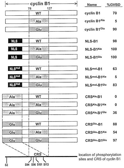
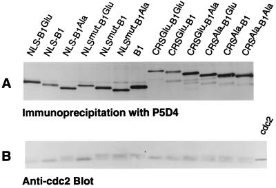
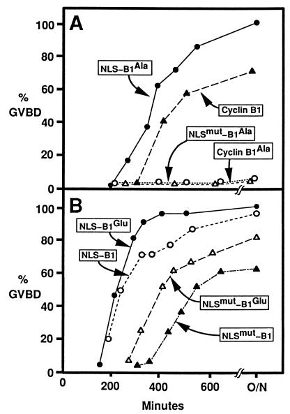
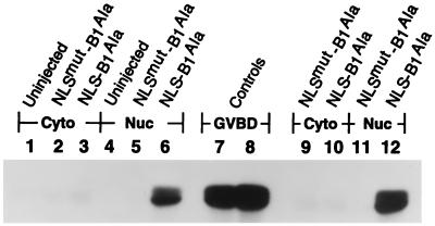
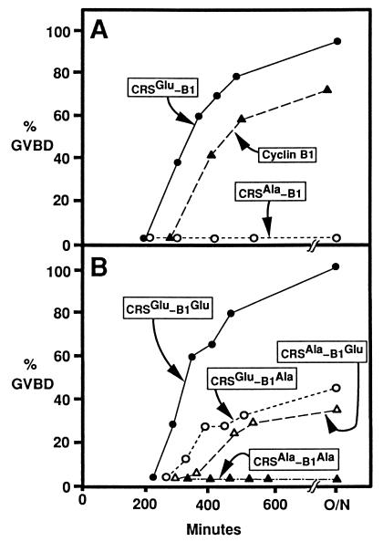
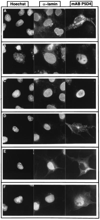
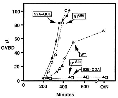
M-phase promoting factor or maturation promoting factor, a key regulator of the G2-->M transition of the cell cycle, is a complex of cdc2 and a B-type cyclin. We have previously shown that Xenopus cyclin B1 has five sites of Ser phosphorylation, four of which map to a recently identified cytoplasmic retention signal (CRS). The CRS appears to be responsible for the cytoplasmic localization of B-type cyclins, although the underlying mechanism is still unclear. Phosphorylation of cyclin B1 is not required for cdc2 binding or cdc2 kinase activity. However, when all of the Ser phosphorylation sites in the CRS are mutated to Ala to abolish phosphorylation, the mutant cyclin B1Ala is inactivated; activity can be enhanced by mutation of these residues to Glu to mimic phosphoserine, suggesting that phosphorylation of cyclin B1 is required for its biological activity. Here we show that biological activity can be restored to cyclin B1Ala by appending either a nuclear localization signal (NLS), or a second CRS domain with the Ser phosphorylation sites mutated to Glu, while fusion of a second CRS domain with the Ser phosphorylation sites mutated to Ala inactivates wild-type cyclin B1. Nuclear histone H1 kinase activity was detected in association with cyclin B1Ala targeted to the nucleus by a wild-type NLS, but not by a mutant NLS. These results demonstrate that nuclear translocation mediates the biological activity of cyclin B1 and suggest that phosphorylation within the CRS domain of cyclin B1 plays a regulatory role in this process. Furthermore, given the similar in vitro substrate specificity of cyclin-dependent kinases, this investigation provides direct evidence for the hypothesis that the control of subcellular localization of cyclins plays a key role in regulating the biological activity of cyclin-dependent kinase-cyclin complexes.


Figures

Figure 1
Figure 1
Structure and activity of cyclin B1-derived fusion proteins. A NLS or nonfunctional mutant NLS (NLSmut) was fused to the N terminus of derivatives of Xenopus cyclin B1. B1 refers to wild-type cyclin B1. B1Ala has Ser → Ala substitutions at residues 94, 96, 101, and 113, and B1Glu has Ser → Glu substitutions at residues 94, 96, 101, and 113. CRSAla and CRSGlu contain Ser → Ala or Ser → Glu substitutions, respectively, at residues 94, 96, 101, and 113. Xenopus oocyte maturation, induced by microinjection of in vitro synthesized RNAs encoding these proteins, was scored as the percentage of microinjected stage VI oocytes that underwent GVBD. The percentage reaching GVBD represents the mean of three independent experiments.
Figure 2
Figure 2
Expression and cdc2 binding of cyclin B1 fusion proteins. Microinjected oocytes were labeled for 5 h as described (13) using 0.5 mCi/ml (1 Ci = 37 GBq) [35S]Met and 0.25 mCi/ml [35S]Cys. (A) Oocytes were lysed and half of the lysate was subjected to immunoprecipitation to recover labeled cyclin proteins using a mAb directed against the epitope tag. Proteins were analyzed by 12.5% SDS/PAGE and detected by fluorography. (B) The other half of each sample was immunoprecipitated as in A, and cdc2 proteins were detected by immunoblotting as described. The last lane in B shows in vitro translated cdc2 as a control.
Figure 3
Figure 3
Kinetics of oocyte maturation in response to cyclin B1 fusion proteins containing a wild-type NLS or NLSmut derived from nucleoplasmin. In vitro-synthesized RNA of each construct was microinjected into a minimum of 20 stage VI oocytes. Oocytes were monitored every 30 min to determine whether they had reached GVBD. (A) NLS–B1Ala, NLSmut–B1Ala, and cyclin B1Ala. (B) NLS–B1, NLS–B1Glu, NLSmut–B1, and NLSmut–B1Glu.
Figure 4
Figure 4
Localization of NLS–B1Ala and NLSmut–B1Ala associated H1 kinase activity. Nuclei were manually isolated at either 2.5 or 4.5 h from oocytes microinjected with in vitro-synthesized RNA encoding NLS–B1Ala and NLSmut–B1Ala. Nuclear and cytoplasmic fractions were then immunoprecipitated using P5D4 antibody to recover the epitope-tagged cyclin B1 fusion proteins, and immunoprecipitates were examined for histone H1 kinase activity, associated with active MPF. Phosphorylated histone H1 was detected by autoradiography after separation by 15% SDS/PAGE. Samples from nuclei (Nuc) or from enucleated oocytes (Cyto) were analyzed at two different time points after microinjection; 2.5 h (lanes 2, 3, 5, and 6) and 4.5 h (lanes 9–12). As a negative control, the cytoplasmic (lane 1) and nuclear (lane 4) fractions from uninjected oocytes were similarly analyzed. For lanes 1–6 and 9–12, each sample corresponds to three nuclei or enucleated oocytes. As a positive control, oocytes that reached GVBD (at ≈5.5 h) after microinjection with RNA encoding NLS–B1Ala were similarly analyzed for histone H1 kinase activity. Lanes 7 and 8 correspond to one-quarter and to one-half of an oocyte, respectively.
Figure 5
Figure 5
Kinetics of oocyte maturation in response to cyclin B1 fusion proteins containing a second CRS appended at the N terminus. Procedures for RNA injections and monitoring oocytes were the same as in Fig. 3. (A) CRSAla–B1 and CRSGlu–B1. (B) CRSAla–B1Ala, CRSAla–B1Glu, CRSGlu–B1Ala, and CRSGlu–B1Glu.
Figure 6
Figure 6
Localization of cyclin B1 mutants in COS-1 cells by indirect immunofluorescence. Hoechst dye 33342 immunofluorescence (Left); α-lamin immunofluorescence (Center); and mAb P5D4 immunofluorescence (Right) to detect cyclin B1 derivatives: cyclin B1Ala (A), cyclin B1Glu (B), NLS–B1Ala (C), NLSmut–B1Ala (D), CRSAla–B1Ala (E), and CRSGlu-B1Glu (F).
Figure 7
Figure 7
Kinetics of oocyte maturation in response to cyclin B1 mutants at phosphorylation sites. Procedures for RNA injections and monitoring oocytes were the same as in Fig. 3.

References

    1. Nigg E A. BioEssays. 1995;17:471–480. - PubMed
    1. Doree M, Galas S. FASEB J. 1994;8:1114–1121. - PubMed
    1. Ducommun B, Brambilla P, Felix M-A, Franza B R, Karsenti E, Draetta G. EMBO J. 1991;10:3311–3319. - PMC - PubMed
    1. Gould K L, Moreno S, Owen D J, Sazer S, Nurse P. EMBO J. 1991;10:3297–3309. - PMC - PubMed
    1. Krek W, Nigg E A. EMBO J. 1991;10:305–316. - PMC - PubMed

Publication types

LinkOut - more resources