

Membrane specific mapping and colocalization of malarial and host skeletal proteins in the Plasmodium falciparum infected erythrocyte by dual-color near-field scanning optical microscopy
- PMID: 9012816
- PMCID: PMC19545
- DOI: 10.1073/pnas.94.2.520
Membrane specific mapping and colocalization of malarial and host skeletal proteins in the Plasmodium falciparum infected erythrocyte by dual-color near-field scanning optical microscopy
Abstract
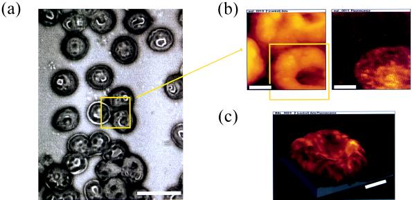
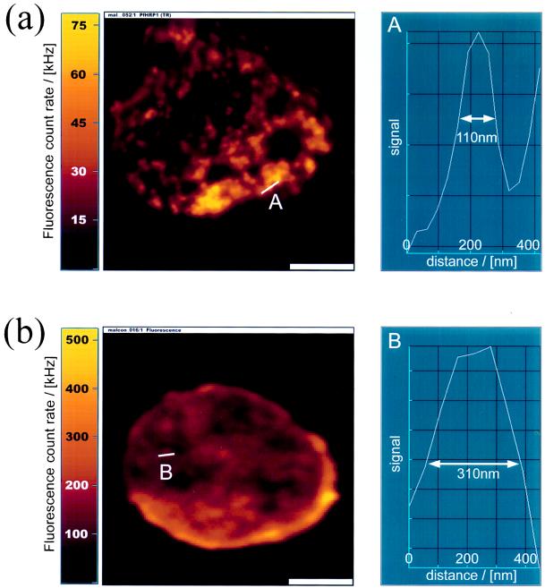
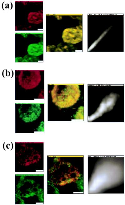
Accurate localization of proteins within the substructure of cells and cellular organelles enables better understanding of structure-function relationships, including elucidation of protein-protein interactions. We describe the use of a near-field scanning optical microscope (NSOM) to simultaneously map and detect colocalized proteins within a cell, with superresolution. The system we elected to study was that of human red blood cells invaded by the human malaria parasite Plasmodium falciparum. During intraerythrocytic growth, the parasite expresses proteins that are transported to the erythrocyte cell membrane. Association of parasite proteins with host skeletal proteins leads to modification of the erythrocyte membrane. We report on colocalization studies of parasite proteins with an erythrocyte skeletal protein. Host and parasite proteins were selectively labeled in indirect immunofluorescence antibody assays. Simultaneous dual-color excitation and detection with NSOM provided fluorescence maps together with topography of the cell membrane with subwavelength (100 nm) resolution. Colocalization studies with laser scanning confocal microscopy provided lower resolution (310 nm) fluorescence maps of cross sections through the cell. Because the two excitation colors shared the exact same near-field aperture, the two fluorescence images were acquired in perfect, pixel-by-pixel registry, free from chromatic aberrations, which contaminate laser scanning confocal microscopy measurements. Colocalization studies of the protein pairs of mature parasite-infected erythrocyte surface antigen (MESA) (parasite)/protein4.1(host) and P. falciparum histidine rich protein (PfHRP1) (parasite)/protein4.1(host) showed good real-space correlation for the MESA/protein4.1 pair, but relatively poor correlation for the PfHRP1/protein4.1 pair. These data imply that NSOM provides high resolution information on in situ interactions between proteins in biological membranes. This method of detecting colocalization of proteins in cellular structures may have general applicability in many areas of current biological research.
Figures
References
-
- Fields S. Methods: Comp Methods Enzymol. 1993;5:116–124.
-
- Smith G P, Scott J K. Methods Enzymol. 1993;217:228–257. - PubMed
-
- Pohl D W, Denk W, Lanz M. Appl Phys Lett. 1984;44:651–653.
-
- Lewis A, Isaacson M, Harootunian A, Murray A. Ultramicroscopy. 1984;13:227–231.
-
- Kirsch A, Meyer C, Jovin T M. In: Proceedings of NATO Advanced Research Workshop: Analytical Use of Fluorescent Probes in Oncology. Kohen E, Hirschberg J G, editors. New York: Plenum; 1996. in press.
Publication types
MeSH terms
Substances
Grants and funding
LinkOut - more resources
Full Text Sources
Other Literature Sources
