. 1997 Jan 21;94(2):657-62.
doi: 10.1073/pnas.94.2.657.

Epidermal growth factor receptor activation induces nuclear targeting of cyclooxygenase-2, basolateral release of prostaglandins, and mitogenesis in polarizing colon cancer cells

Affiliations

Epidermal growth factor receptor activation induces nuclear targeting of cyclooxygenase-2, basolateral release of prostaglandins, and mitogenesis in polarizing colon cancer cells

R J Coffey et al. Proc Natl Acad Sci U S A. .

Abstract

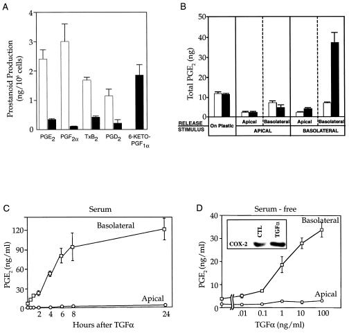
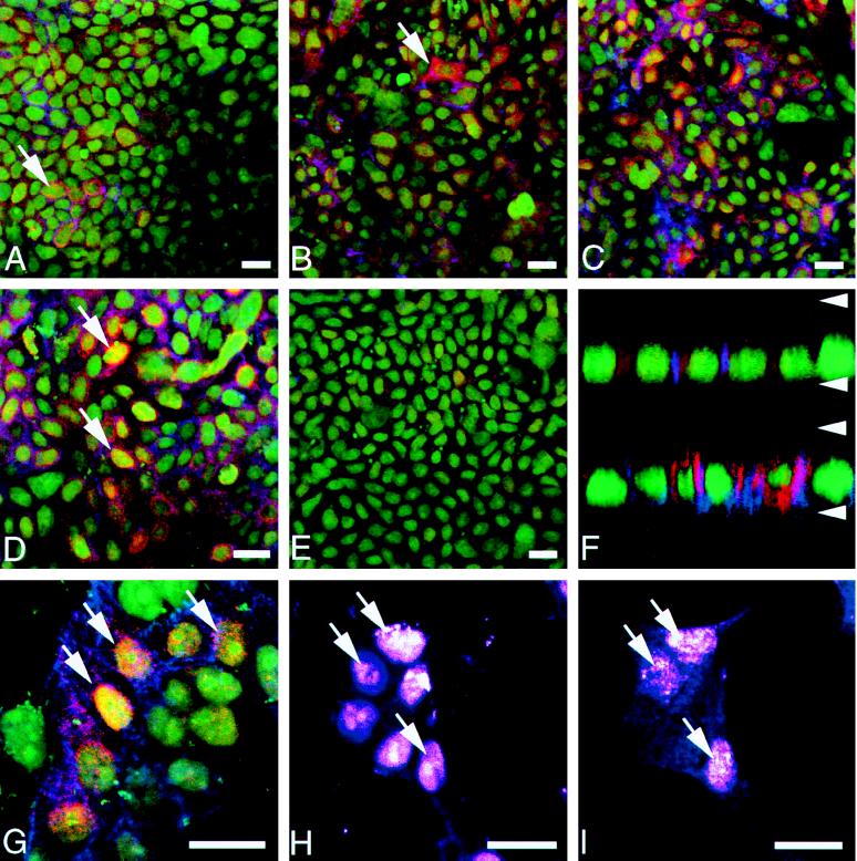
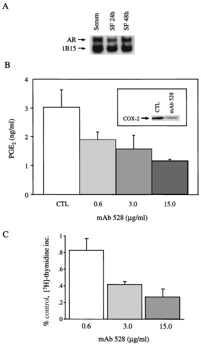
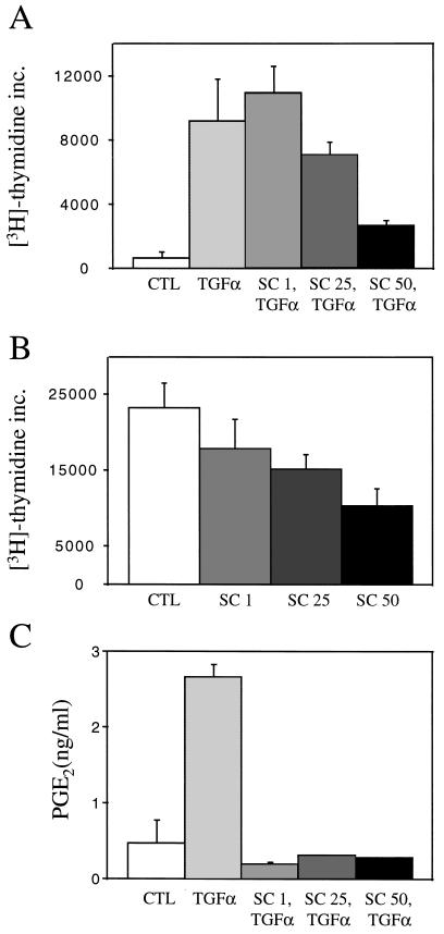
Nonsteroidal antiinflammatory drugs reduce the risk of colon cancer, possibly via cyclooxygenase (COX) inhibition. The growth factor-inducible COX-2, which is overexpressed in neoplastic colonic tissue, is an attractive target to mediate this effect. Herein we have exploited the ability of a human colon cancer cell line, HCA-7 Colony 29, to polarize when cultured on Transwell (Costar) filters to study COX-2 production and the vectorial release of prostaglandins (PGs). Administration of type alpha transforming growth factor to the basolateral compartment, in which the epidermal growth factor receptor (EGFR) resides, results in a marked induction of COX-2 immunoreactivity at the base of the cells and the unexpected appearance of COX-2 in the nucleus. The increase in COX-2 protein is associated with a dose- and time-dependent increase in PG levels in the basolateral, but not apical, medium. Amphiregulin is the most abundantly expressed EGFR ligand in these cells, and the protein is present at the basolateral surface. EGFR blockade reduces baseline COX-2 immunoreactivity, PG levels, and mitogenesis in a concentration-dependent manner. Two specific COX-2 inhibitors, SC-58125 and NS 398, also, in a dose-dependent manner, attenuate baseline and type alpha transforming growth factor-stimulated mitogenesis, although PG levels are decreased > 90% at all concentrations of inhibitor tested. These findings show that activation of the EGFR stimulates COX-2 production and its translocation to the nucleus, vectorial release of PGs, and mitogenesis in polarized HCA-7 Colony 29 cells.


Figures

Figure 1
Figure 1
Eicosanoid production by HCA-7 and RIE-1 cells. (A) Rapidly growing HCA-7 (open bars) and RIE-1 (filled bars) cells cultured on plastic in 10% fetal calf serum were stimulated by AA (20 μM) for 30 min before harvest, and prostanoid levels in the medium were quantified by gas chromatography/negative ion chemical ionization mass spectrometry using stable isotope dilution techniques (25). (B) HCA-7 cells, cultured as a flat monolayer on plastic or as a polarizing monolayer on Transwell filters in 10% fetal calf serum, were exposed to TGFα (100 ng/ml; filled bars) or diluent (open bars). A 30-min pulse of AA (20 μM) was given 7.5 h later, and PGE2 in the medium was measured. (C) TGFα (10 ng/ml) was added to the basal medium of HCA-7 cells cultured in 10% fetal calf serum on Transwell filters. Thirty minutes before each time point, AA (20 μM) was added and PGE2 was measured in the apical or basolateral medium. (D) TGFα at varying concentrations was added to the basal medium of HCA-7 cells that had been serum-free for 48 h. A 30-min pulse of AA (20 μM) was given 7.5 h later, and PGE2 in the apical or basolateral medium was measured. (Inset) COX-2 immunoblot of HCA-7 cells grown on Transwell filters that had been serum-free for 48 h and then treated with TGFα (10 ng/ml) for 24 h. TGFα stimulated PGE2 release, following addition of exogenous AA (20 μM) and endogenous stores of AA.
Figure 2
Figure 2
Distribution of COX-2 immunofluorescence in HCA-7 cells imaged with confocal microscopy. Both confluent (AF) and sparsely plated cells (GI) were fixed and labeled with rabbit anti-COX-2 (red), mAb to AR (blue), and a nuclear fluorochrome (green). (A) Overview of cells with serum withdrawn 24 h before fixation. Note that a few small clusters (arrow) exhibit constitutive COX-2 and AR fluorescent signals. (B) Four hours after the addition of TGFα to culture medium, COX-2 immunofluorescence has increased in some cells with a few cells with high levels (arrow). (C) After 8 h of TGFα treatment, HCA-7 cell culture exhibits many cells with significant levels of COX-2 immunofluorescence. The AR signal has also increased in this cell population, but not necessarily in the same cells with increased COX-2. (D) Twenty-four hours after the addition of TGFα, most of the cells display a bright cytoplasmic COX-2 signal, which is highest in the perinuclear region; however, a few cells exhibit both cytoplasmic and nuclear COX-2 immunofluorescence (arrows). (E) HCA-7 cells pretreated with 15 μg/ml mAb 528 for 24 h before immunostaining results in diminished COX-2 signal. (F) Confocal xz scan analysis of confluent HCA-7 cells either serum-deprived (upper) or TGFα-treated for 24 h (lower) shows significant enhancement of perinuclear COX-2 signal following treatment with TGFα. Arrowheads mark the approximate positions of base and apex of cells with a distance of 8.5 μm between them. (G) Higher magnification of sparsely plated cells 24 h after TGFα addition reveals that many cells display enhanced COX-2 and AR expression. Cells with strong nuclear COX-2 immunoreactivity (arrow) often exhibit striking perinuclear COX-2 signal as well. (H) Optical section through sparsely plated cells stained with nuclear fluorochrome YO-PRO1 (arrows) and (I) for COX-2 at same optical plane, demonstrating nuclear COX-2 immunoreactivity (arrows). (Bars = 10 μm.)
Figure 3
Figure 3
EGFR-regulated COX-2 production, basolateral release of PGs and mitogenesis in polarized HCA-7 cells. (A) Northern blot of HCA-7 cells grown on Transwell filters in 10% fetal calf serum and then kept in serum-free medium for 24 and 48 h. Two micrograms of poly(A) RNA was probed with cRNA probes for AR and constitutively expressed 1B15. (B) HCA-7 cells, grown on Transwell filters and maintained serum-free for 24 h, were exposed to increasing concentrations of mAb 528, and PGE2 levels in the basolateral medium were measured by mass spectrometry. (Inset) COX-2 immunoblot of HCA-7 cells under identical starting conditions as control cells that were then treated with mAb 528 (3 μg/ml) for 24 h. (C) Under identical experimental conditions as in B, a 3-h pulse of [3H]thymidine (1 μCi/ml) was administered 21 h after treatment with mAb 528 and trichloroacetic acid-precipitable counts were determined. All experiments were performed in triplicate and repeated at least twice. The mitogenic data were expressed as percent of control. Error bars represent standard deviation.
Figure 4
Figure 4
COX-2 inhibition decreases TGFα-stimulated and baseline mitogenesis in polarized HCA-7 cells. (A) HCA-7 cells maintained 48 h serum-free on Transwell filters were pretreated for 1 h with increasing concentrations of SC-58125, and then TGFα (10 ng/ml) was administered. (B) HCA-7 cells cultured on Transwell filters in 10% serum were treated with increasing concentrations of SC-58125. In both experiments, a 3-h pulse of [3H]thymidine (1 μCi/ml) was performed 21 h after administration of the agents and trichloroacetic acid-precipitable counts were determined. (C) Under identical conditions as in A, a 30-min pulse of AA (20 μM) was administered 7.5 h after TGFα and PGE2 levels in the basolateral medium were determined by mass spectrometry. All experiments were performed in triplicate and repeated at least three times. Error bars represent standard deviation.

References

    1. Eberhart C, DuBois R. Gastroenterology. 1995;109:285–301. - PubMed
    1. Cuthbert A, Halushka P, Margolius H, Spayne J. Br J Pharmacol. 1984;82:597–607. - PMC - PubMed
    1. Bito L, Wallenstein M, Baroody R. Adv Prostaglandin Thromboxane Res. 1976;1:297–303. - PubMed
    1. Garcia-Perez A, Smith W. J Clin Invest. 1984;74:63–74. - PMC - PubMed
    1. Cortizo A, Besterman J, Leitner P, Chandrabose K. Prostaglandins. 1992;44:357–371. - PubMed

Publication types

MeSH terms

Substances