. 1997 Jan 21;94(2):663-8.
doi: 10.1073/pnas.94.2.663.

Constitutive production and thrombin-induced release of vascular endothelial growth factor by human megakaryocytes and platelets

Affiliations

Constitutive production and thrombin-induced release of vascular endothelial growth factor by human megakaryocytes and platelets

R Möhle et al. Proc Natl Acad Sci U S A. .

Abstract

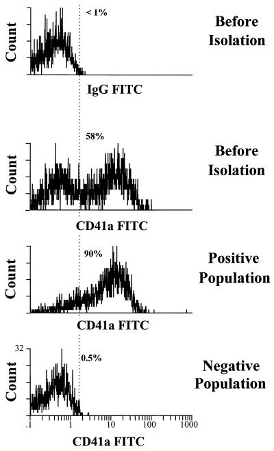
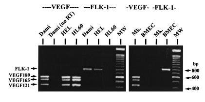
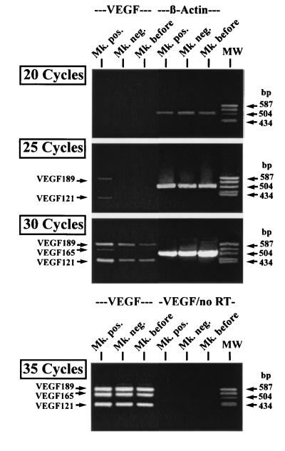
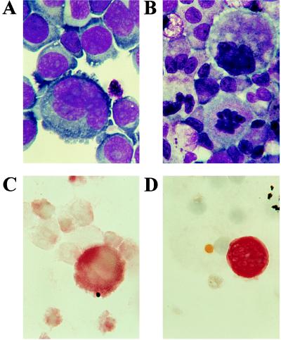
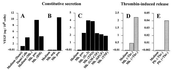
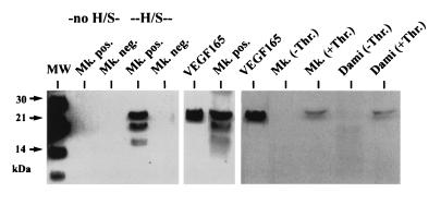
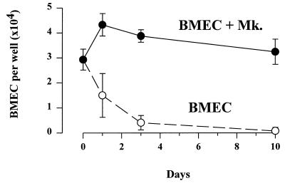
We have shown that coculture of bone marrow microvascular endothelial cells with hematopoietic progenitor cells results in proliferation and differentiation of megakaryocytes. In these long-term cultures, bone marrow microvascular endothelial cell monolayers maintain their cellular integrity in the absence of exogenous endothelial growth factors. Because this interaction may involve paracrine secretion of cytokines, we evaluated megakaryocytic cells for secretion of cytokines, we evaluated megakaryocytic cells for secretion of vascular endothelial growth factor (VEGF). Megakaryocytes (CD41a+) were generated by ex vivo expansion of hematopoietic progenitor cells with kit-ligand and thrombopoietin for 10 days and further purified with immunomagnetic microbeads. Using reverse transcription-PCR, we showed that megakaryocytic cell lines (Dami, HEL) and purified megakaryocytes expressed mRNA of the three VEGF isoforms (121, 165, and 189 amino acids). Large quantities of VEGF (> 1 ng/10(6) cells/3 days) were detected in the supernatant of Dami cells, ex vivo-generated megakaryocytes, and CD41a+ cells isolated from bone marrow. The constitutive secretion of VEGF by CD41a+ cells was stimulated by growth factors of the megakaryocytic lineage (interleukin 3, thrombopoietin). Western blotting of heparin-Sepharose-enriched supernatant mainly detected the isoform VEGF165. In addition, immunohistochemistry showed intracytoplasmic VEGF in polyploid megakaryocytes. Thrombin stimulation of megakaryocytes and platelets resulted in rapid release of VEGF within 30 min. We conclude that human megakaryocytes produce and secrete VEGF in an inducible manner. Within the bone marrow microenvironment, VEGF secreted by megakaryocytes may contribute to the proliferation of endothelial cells. VEGF delivered to sites of vascular injury by activated platelets may initiate angiogenesis.


Figures

Figure 1
Figure 1
Isolation of CD41a+ megakaryocytic cells. Megakaryocytes were separated after labeling with anti-CD41a–FITC antibody and anti-FITC immunomagnetic microbeads. Cells before isolation, the positive and negative cell fraction, were analyzed by flow cytometry. While the positive fraction contained 90% CD41a+ cells, virtually no megakaryocytic cells were found in the negative population (0.5%).
Figure 2
Figure 2
RT-PCR of VEGF and FLK-1 mRNA expression. The megakaryocytic cell lines Dami and HEL, as well as ex vivo-generated megakaryocytes (Mk.), expressed mRNA of the three VEGF isoforms (VEGF189, VEGF165, and VEGF121). Only the malignant cell lines simultaneously expressed FLK-1 mRNA. RNA from Dami cells analyzed without reverse transcription (no RT) served as a negative control, and RNA from the cell line HL-60 as a positive control for VEGF expression. Endothelial cells (BMEC) as a positive control expressed only FLK-1.
Figure 3
Figure 3
Semiquantitative RT-PCR of VEGF mRNA expression. Equal amounts of cDNA from cells before isolation (Mk. before), CD41a+ (Mk. pos.), and CD41a (Mk. neg.) cells were amplified by RT-PCR for VEGF and β-actin expression using different numbers of amplification cycles. VEGF121 and VEGF189 were detected after 25 PCR cycles only in CD41a+ cells. Furthermore, VEGF165 was detected after 30 cycles only in these cells. In contrast, no difference in the β-actin expression was observed. After 35 PCR cycles, PCR products corresponding to the three VEGF isoforms could also be amplified from CD41a and unseparated cells. As a negative control, RNA without RT was also subjected to 35 cycles of PCR amplification (VEGF/no RT).
Figure 4
Figure 4
Immunohistochemistry of VEGF expression. Cytospin preparations of Dami cells and megakaryocytes were stained with Wright–Giemsa (A and B, respectively) and analyzed with immunohistochemistry for VEGF expression (C and D). The megakaryocytic cell line Dami was positive for VEGF (C), particularly with increasing cell size. Among the ex vivo-generated megakaryocytic cells, only highly polyploid, large megakaryocytes stained positive for VEGF (D). (×1000.)
Figure 5
Figure 5
(A and B) Constitutive secretion of VEGF. During 72 h of incubation (A), Dami-cells, ex vivo-generated (Mk. pos.) and bone marrow-derived megakaryocytes (Mk. BM) released VEGF into the supernatant. Medium not conditioned by cells served as control (Medium). Because the Dami cell line was cultured in serum-containing medium, VEGF was also detected in the respective control sample (Medium Dami). In a second experiment (B), secretion of VEGF by CD41a+ megakaryocytes was compared with CD41 cells. In contrast to CD41a+ cells (Mk. pos.), the amount of VEGF secreted by CD41a cells during 72 h (Mk. neg.) was below detection limit. (C) Addition of IL-3 and TPO resulted in an increased secretion of VEGF by isolated CD41a+ cells during an incubation period of 24 h. GM-CSF, bFGF, or tumor necrosis factor α (TNF-α) had no effect or decreased VEGF secretion. (D and E) Thrombin-induced release of VEGF. Megakaryocytes and platelets were isolated, washed, and resuspended in serum-free medium. Thirty minutes after incubation with thrombin at a concentration of 1 unit/ml, VEGF was detected in the supernatant from megakaryocytes (Mk. +Thr.) (D) and platelets (Plt. +Thr). (E), compared with medium from unstimulated cells (Mk. −Thr., Plt. −Thr.).
Figure 6
Figure 6
Western blotting of conditioned medium from megakaryocytic cells. Without heparin-Sepharose enrichment (no H/S), no VEGF could be detected in the conditioned medium (72-h incubation) of CD41a+ (Mk. pos.) and CD41a (Mk. neg.) cells. Two bands at 21–22 and 17–18 kDa were visible after enrichment of supernatant from CD41a+ cells with heparin-Sepharose (H/S), corresponding to glycolysated and nonglycolysated VEGF165, while no detectable protein was found in the conditioned medium from CD41a cells. The identity of the bands was confirmed by the positive control (recombinant VEGF165). Thirty minutes after stimulation with thrombin, VEGF165 was also detectable in the supernatant from ex vivo-generated megakaryocytes (Mk. +Thr.) and Dami-cells (Dami +Thr.) after enrichment with H/S, in contrast to unstimulated cells (−Thr.).
Figure 7
Figure 7
Coculture of BMEC and megakaryocytes. In coculture with megakaryocytes, BMEC monolayers could be maintained for longer than 1 week in serum-free medium (BMEC +Mk). In contrast, the number of BMEC per well (mean ± SD) rapidly dropped without coculture (BMEC).

References

    1. Rafii S, Shapiro F, Pettengell R, Ferris B, Nachman R L, Moore M A S, Asch A S. Blood. 1995;86:3353–3363. - PubMed
    1. Tavassoli M, Aoki M. Blood Cells. 1989;15:59–72. - PubMed
    1. Wickenhauser C, Lorenzen J, Thiele J, Hillienhof A, Jungheim K, Schmitz B, Hansmann M, Fischer R. Blood. 1995;85:685–691. - PubMed
    1. Jones C L A, Witte D P, Feller M J, Fugman D A, Dorn G W, Liebermann M A. Biochim Biophys Acta. 1992;1136:272–282. - PubMed
    1. Connolly D T, Heuvelman D M, Nelson R, Olander J V, Eppley B L, Delfino J J, Siegel N R, Leimgruber R M, Feder J. J Clin Invest. 1989;84:1470–1478. - PMC - PubMed

Publication types

MeSH terms

Substances

LinkOut - more resources