. 1997 Jan 21;94(2):724-8.
doi: 10.1073/pnas.94.2.724.

Dendritic calcium conductances generate high-frequency oscillation in thalamocortical neurons

Affiliations

Dendritic calcium conductances generate high-frequency oscillation in thalamocortical neurons

C Pedroarena et al. Proc Natl Acad Sci U S A. .

Abstract

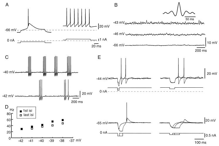
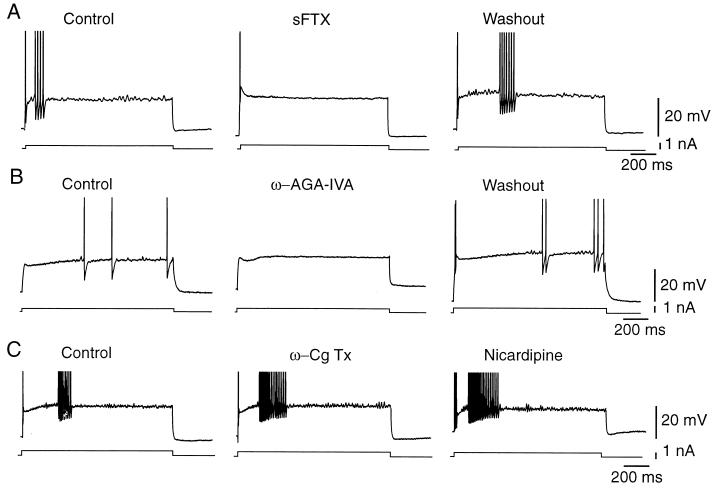
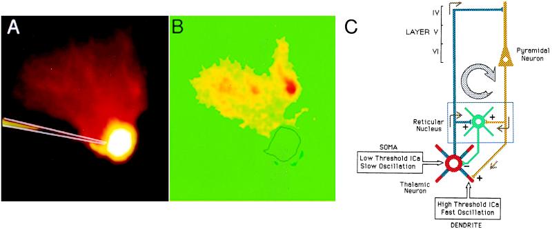
Cortical-projecting thalamic neurons, in guinea pig brain slices, display high-frequency membrane potential oscillations (20-80 Hz), when their somata are depolarized beyond -45 mV. These oscillations, preferentially located at dendritic sites, are supported by the activation of P/Q type calcium channels, as opposed to the expected persistent sodium conductance responsible for such rhythmic behavior in other central neurons. Short hyperpolarizing pulses reset the phase and transiently increase the amplitude of these oscillations. This intrinsic thalamic electroresponsiveness may serve as a cellular-based temporal binding mechanism that sharpens the temporal coincidence of cortical-feedback synaptic inputs, known to distribute at remote dendritic sites on thalamic neurons.


Figures

Figure 1
Figure 1
Fast oscillations in TCNs. (A) Direct activation of a thalamic cell from two levels of membrane potential. The labeled −66-mV broken line indicates the resting membrane potential. (B) In the same cell, subthreshold oscillations at two different levels of membrane potential induced by outward dc injection. The dominant frequency at −43 mV was 37.5 Hz (see Inset, autocorrelogram). (C) High-frequency trains of spikes induced by increasing dc injection. Note that the spikes are triggered at the same frequency as the subthreshold oscillations. (Filtering of the traces at 2 kHz reduced the amplitude of the spikes.) (D) Corresponding graph of discharge frequency vs. membrane potential. The rate of discharges are in the gamma band. isi, inter-spike interval. (E) Oscillations are reset (upper traces) by brief hyperpolarizations induced by the injection of short-duration inward current pulses. (Lower traces) Low-threshold spikes can be induced by outward current pulses of longer duration or higher amplitudes than those in upper traces, even from resting membrane potential (−65 mV).
Figure 2
Figure 2
Ionic mechanisms of the fast subthreshold membrane potential oscillation in thalamocortical cells. (A) TTX did not block oscillation, but increased voltage threshold (Insets, autocorrelograms). (B) Plot of voltage responses to outward and inward current pulses from −60 mV, before and after TTX. Following TTX, the effective depolarization decreased (Inset) and a strong outward rectification for membrane potentials positive to −50 mV became evident. (C) NaCl equimolar substitution with choline chloride did not block oscillation. (D) CdCl2 (up to 0.2 mM) blocked the fast subthreshold membrane potential oscillations but not the low-threshold spikes (arrows). (E) Fast subthreshold oscillations became more evident after equimolar replacement of CaCl2 by BaCl2.
Figure 3
Figure 3
Calcium channel blockers and the oscillatory activity of thalamic cells. (A) sFTX (1 mM) to the recording chamber completely blocked the fast subthreshold oscillations and reversed after 15 min washout. (B) Peptidyl toxin from A. aperta (100 nM) also reversibly blocked the fast oscillation. (C) ω-conotoxin (10 μM) or nicardipine (2 μM) had no affect on fast oscillatory activity.
Figure 4
Figure 4
Illustration of the [Ca]i location during depolarization of a thalamic cell to the level of fast membrane potential oscillations. (A) Fura-2 control fluorescent image of an iontophoretically filled neuron. (B) [Ca]i image obtained from subtracting background fluorescence from recordings acquired during a depolarizing step to membrane potential level positive to the threshold for oscillation. Note the increase in [Ca]i at dendritic level. (C) Diagram of the proposed thalamocortical neuronal circuit depicting the somatic and dendritic calcium conductances in thalamic neurons. A thalamic projection neuron is shown synapsing on a pyramidal cell at layer IV. The return pathway to the thalamus terminates mostly on the dendritic compartments of TCNs, where dendritic high-threshold calcium-dependent oscillations are observed (red). The corticothalamic input gives collaterals to the reticular nucleus (green) reinforcing thalamocortical resonance by inhibitory reset.

References

    1. Jahnsen H, Llinás R. J Physiol (London) 1984;349:105–226. - PubMed
    1. Jahnsen H, Llinás R. J Physiol (London) 1984;349:227–247. - PMC - PubMed
    1. Leresche N, Lightower S, Soltesz I, Jassik-Gerschenfeldt D, Crunelli D. J Physiol (London) 1991;441:155–174. - PMC - PubMed
    1. McCormick D A, Pape H C. J Physiol (London) 1990;431:291–318. - PMC - PubMed
    1. Steriade M, Curró Dossi R, Contreras D. Neuroscience. 1993;56:1–9. - PubMed

Publication types

LinkOut - more resources