. 1997 Jan 27;136(2):251-69.
doi: 10.1083/jcb.136.2.251.

Biogenesis of the Saccharomyces cerevisiae mating pheromone a-factor

Affiliations

Biogenesis of the Saccharomyces cerevisiae mating pheromone a-factor

P Chen et al. J Cell Biol. .

Abstract

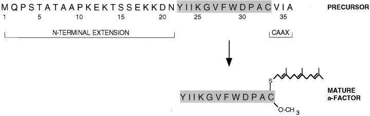
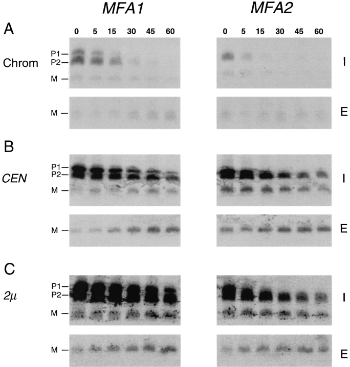
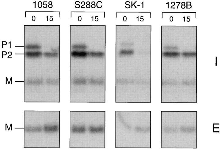
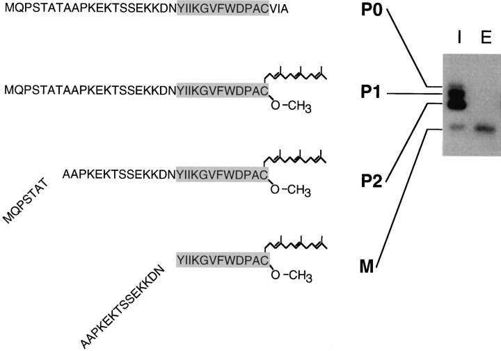
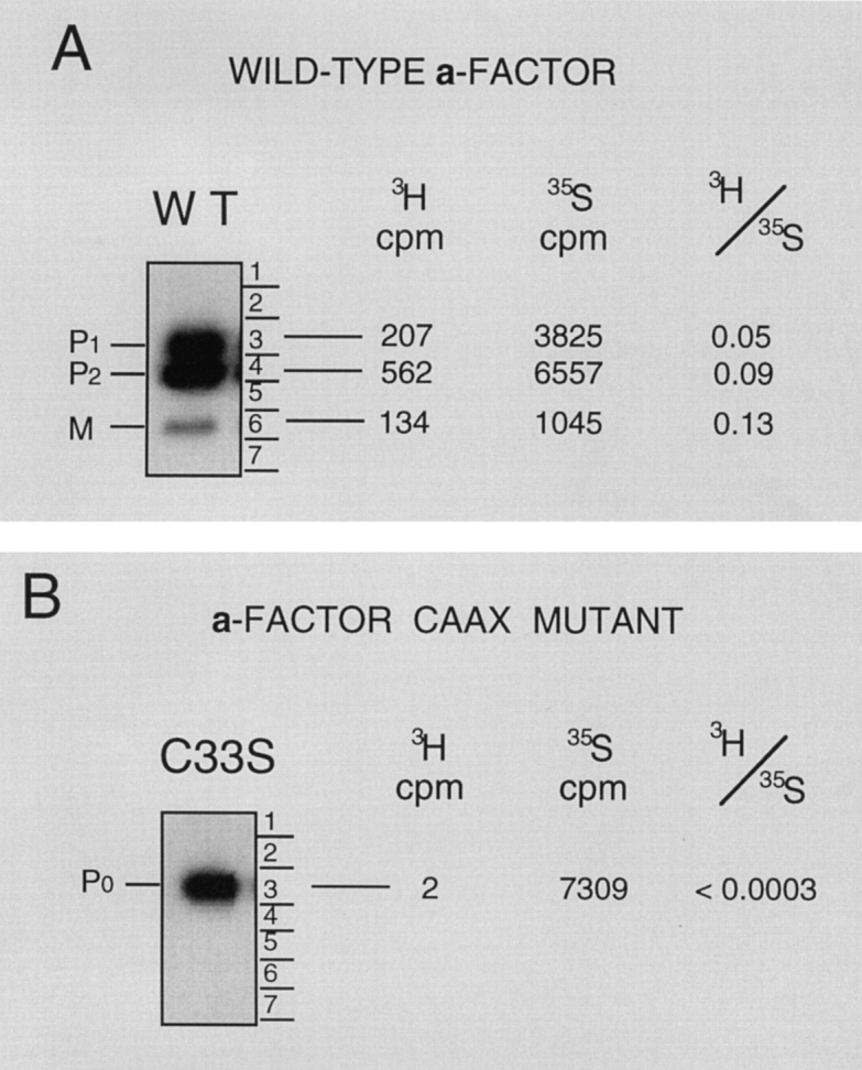
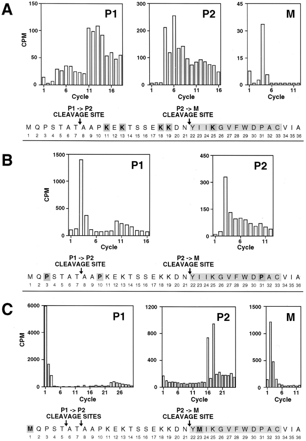
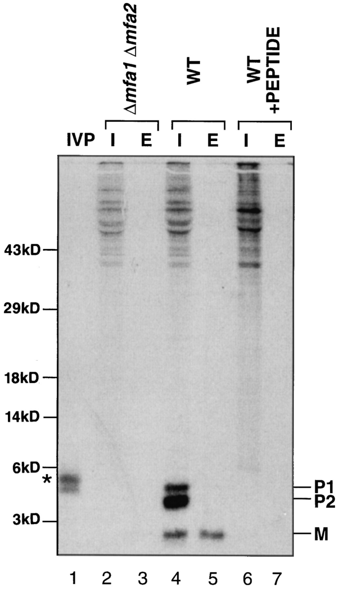
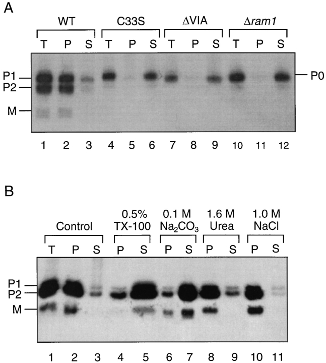
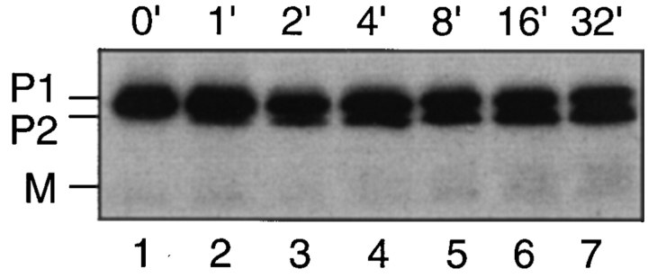
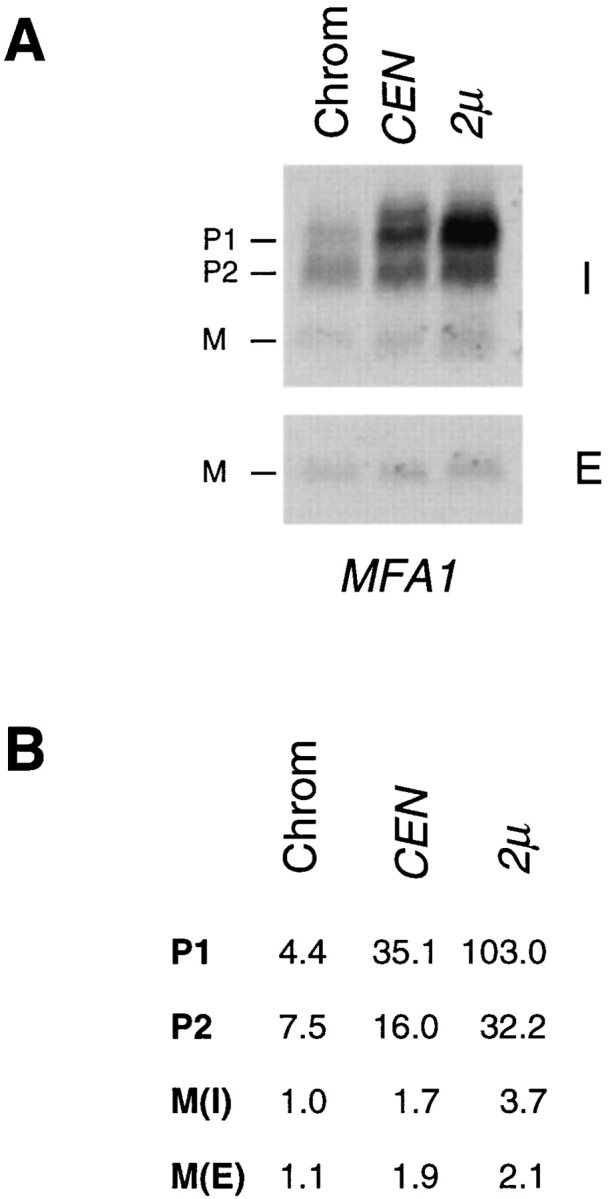
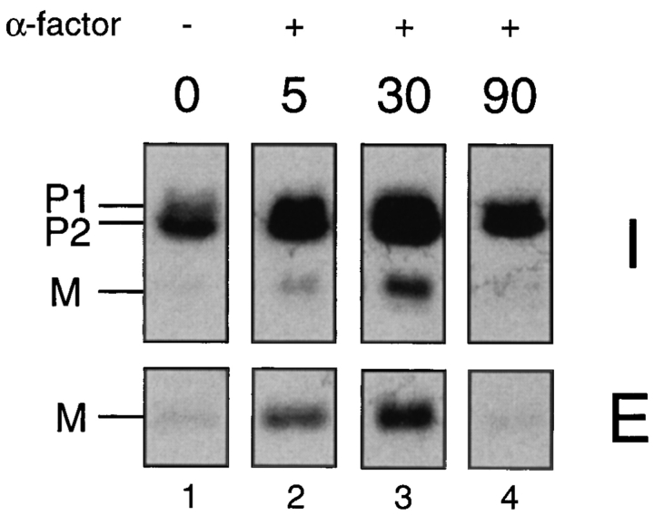
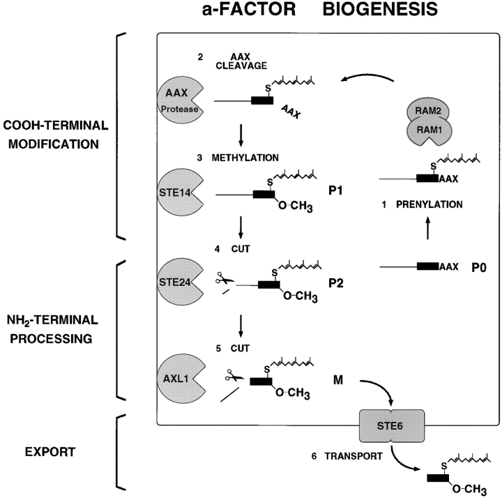
The Saccharomyces cerevisiae mating pheromone a-factor is a prenylated and carboxyl methylated extracellular peptide signaling molecule. Biogenesis of the a-factor precursor proceeds via a distinctive multistep pathway that involves COOH-terminal modification. NH2-terminal proteolysis, and a nonclassical export mechanism. In this study, we examine the formation and fate of a-factor biosynthetic intermediates to more precisely define the events that occur during a-factor biogenesis. We have identified four distinct a-factor biosynthetic intermediates (P0, P1, P2, and M) by metabolic labeling, immunoprecipitation, and SDS-PAGE. We determined the biochemical composition of each by defining their NH2-terminal amino acid and COOH-terminal modification status. Unexpectedly, we discovered that not one, but two NH2-terminal cleavage steps occur during the biogenesis of a-factor. In addition, we have shown that COOH-terminal prenylation is required for the NH2-terminal processing of a-factor and that all the prenylated a-factor intermediates (P1, P2, and M) are membrane bound, suggesting that many steps of a-factor biogenesis occur in association with membranes. We also observed that although the biogenesis of a-factor is a rapid process, it is inherently inefficient, perhaps reflecting the potential for regulation. Previous studies have identified gene products that participate in the COOH-terminal modification (Ram1p, Ram2p, Ste14p), NH2-terminal processing (Ste24p, Axl1p), and export (Ste6p) of a-factor. The intermediates defined in the present study are discussed in the context of these biogenesis components to formulate an overall model for the pathway of a-factor biogenesis.


Figures

Figure 1
Figure 1
Structure of precursor and mature forms of a-factor encoded by MFA1. The a-factor precursor encoded by MFA1 is shown with the NH2-terminal extension, COOH-terminal CAAX motif, and mature portion (shaded gray) indicated. Every fifth residue is numbered. Mature a-factor derived from this precursor is modified on its COOH-terminal cysteine residue by a farnesyl moiety and a carboxyl methyl group, as indicated.
Figure 8
Figure 8
Kinetic analysis of a-factor biogenesis in strains expressing MFA1 or MFA2 at low (chromosomal), intermediate (CEN), or high () levels. Cells were pulse labeled with [35S]cysteine for 5 min, and the label was chased for the times indicated. Intracellular (I) and extracellular (E) fractions were prepared, and proteins were subjected to immunoprecipitation, SDS- PAGE, and exposed to a Phosphorimager screen for 24 h. The P1, P2, and M species are indicated. A constant gray scale value is used for all the scans shown, so that the relative level of a-factor species in each strain can be qualitatively compared by direct visual inspection. The strains examined here express MFA1 or MFA2 only, from the chromosome (SM2892 and SM3019), a CEN plasmid (SM2891 and SM3020), and a plasmid (SM2893 or SM3021).
Figure 9
Figure 9
Comparison of the overall a-factor biogenesis pattern in yeast strains derived from distinct genetic lineages. Strains SM1058, S288C, SK-1, and Σ1278B were pulse labeled with [35S]cysteine for 5 min. Intracellular (I) and extracellular (E) a-factor species were immunoprecipitated immediately or after a 15-min chase and analyzed by SDS-PAGE and autoradiography.
Figure 5
Figure 5
The deduced structure of a-factor intermediates. The SDS-PAGE analysis of immunoprecipitated intracellular (I) and extracellular (E) species of a-factor present after a brief (5min) pulse labeling is shown (right). The deduced structure of each a-factor biosynthetic intermediate is indicated (left), based on the results from the carboxyl methylation assay (Fig. 3) and the radiolabeled peptide sequence analysis (Fig. 4). The strain labeled is SM1710, which contains pSM233 (CEN MFA1).
Figure 3
Figure 3
Carboxyl methylation assay to determine the COOHterminal modification status of a-factor biosynthetic intermediates. The methylation status of a-factor biosynthetic intermediates was determined in a strain containing a wild-type MFA1 plasmid (A, SM1585) or a plasmid with the a-factor CAAX mutation mfa1-C33S (B, SM1682), in which prenylation and subsequent modification of a-factor is prevented. Cells were double labeled with [35S]cysteine and [3H-methyl]AdoMet (A) or with Trans35-label and [3H-methyl]AdoMet (B), respectively. Cell extracts were prepared, proteins were immunoprecipitated with a-factor antibodies, separated by 16% SDS-PAGE, and the dried gel was subjected to autoradiography to detect the indicated intracellular a-factor biosynthetic intermediates. Each a-factor intermediate was excised from the gel, and the relative degree of carboxyl methylation was determined. Incorporation of 3H into methyl esters was determined by subjecting the excised gel slice to the vapor phase equilibrium assay in which 3H-labeled methyl ester groups are converted to [3H]methanol counts. Total 35S incorporation was determined by solubilization of the gel slice. The 3H/35S ratio determined by these two tests is shown.
Figure 4
Figure 4
Radiolabeled peptide sequence analysis to establish the NH2-terminal processing status of intracellular species of a-factor. The a-factor species in immunoprecipitates from cells labeled with [3H]lysine (A), [3H]proline (B), or Trans35S label (methionine:cysteine = 20:1) (C) were separated by 16% SDS-PAGE and transferred onto polyvinyldifluoride membrane. The intracellular a-factor intermediates P1, P2, and M were visualized by autoradiography, excised, and subjected to Edman degradation analysis. The radioactive material from each cycle was quantitated by scintillation counting as shown in the bar graph. Within the a-factor sequence, the radiolabeled amino acid used for labeling is shaded darkly, and the deduced P1→ P2 and P2→ M cleavage sites are indicated. The strain labeled in A and B is SM1762, which carries the wild-type a-factor plasmid pSM463 (2μ MFA1). The strain labeled in C is SM1932, which harbors an a-factor substitution mutant plasmid, pSM490 (2μ mfa1-I23M).
Figure 2
Figure 2
Identification of a-factor biosynthetic intermediates. Cells were labeled with [35S]cysteine under steady-state conditions and intracellular (I) and extracellular (E) extracts were prepared. Proteins were immunoprecipitated with a-factor antiserum (Ab-9-137), separated by 16% SDS-PAGE, and visualized by autoradiography, as described in Materials and Methods. The in vitro precursor (IVP; lane 1) was generated by transcription and translation of MFA1 from pSM118. Strains are transformants of an a-factor deletion strain (Δmfa1 Δmfa2) and contain either no plasmid (lanes 2 and 3; SM1458) or an a-factor plasmid, pSM464 (CEN MFA1; lanes 4–7; SM1829). In the peptide competition experiment (lanes 6 and 7), the labeled extracts are the same as in lanes 4 and 5, respectively, except that a 50-fold excess of cold synthetic dodecapeptide (YIIKGVFWDPAC) corresponding to mature a-factor was added before immunoprecipitation. The a-factor precursor species are designated P1 and P2, and the band corresponding to mature a-factor is designated M. Molecular mass markers are indicated. The band in lane 1 just above P1, marked by an asterisk, most likely represents the unmodified a-factor precursor P0.
Figure 6
Figure 6
Fractionation and solubilization properties of intracellular a-factor. In A, cells were labeled with [35S]cysteine for 5 min and lysates were prepared. The total cellular lysate (T) was separated into particulate (P) and soluble (S) fractions by centrifugation at 100,000 g for 1 h at 4°C. The a-factor species were immunoprecipitated and subjected to SDS-PAGE analysis. Strains examined in lanes 1–9 (SM1585, SM1682, and SM1680) carry either a wild-type or mutant MFA1 plasmid, as indicated. Strain SM1865, examined in lanes 10–12, is a Δram1 mutant. In B, a strain containing a wild-type MFA1 plasmid (SM1762) was labeled with [35S]cysteine for 5 min. The total lysate (T) was subjected to the indicated treatments or to no treatment (control, lanes 1–3) and subsequently separated into particulate (P) and soluble (S) fractions by centrifugation at 100,000 g for 1 h at 4°C. The a-factor intermediates were immunoprecipitated and analyzed by SDSPAGE and autoradiography.
Figure 7
Figure 7
Kinetic analysis demonstrating P1→ P2 conversion of the a-factor precursor. Strain SM1762, which carries pSM463 (2μ MFA1), was pulse labeled with [35S]cysteine for 30 s, chased for the indicated time points (in minutes), and extracts were prepared. The intracellular fraction was subjected to immunoprecipitation, SDS-PAGE, and autoradiography.
Figure 10
Figure 10
Comparison of the a-factor biosynthesis profile in strains expressing MFA1 at low (chromosomal), intermediate (CEN), or high () levels. To quantitate the total amount of precursor plus mature a-factor synthesized in each of the MFA1bearing strains shown in Fig. 8, and to determine the relative amounts of each a-factor intermediate, the intracellular immunoprecipitates from the 0-min chase time point and the extracellular immunoprecipitates from the 15-min chase point shown in Fig. 8 were electrophoresed side by side and quantitated by Phosphorimager analysis. A shows the Phosphorimager scan. B shows the quantitation of a-factor intermediates from A. Values are expressed relative to chromosomal intracellular mature a-factor, M(I), which is set at 1.0. M(E) refers to extracellular mature a-factor. These data illustrate two points: the low level of mature (M(I) + M(E)) relative to precursor species, indicative of the low efficiency of P2→ M processing, and the buildup of P1 when a-factor is expressed at very high levels.
Figure 11
Figure 11
Examination of a-factor biogenesis after α-factor induction. Strain SM1227 (MFA1 mfa2::URA3) was preincubated with 25 μM α-factor (except for the 0-min sample). At the indicated times, which represent minutes after the addition of pheromone, cells were pulse labeled with [35S]cysteine for 5 min, and the label was chased for 45 min. (For the 0-min control sample, the pulse chase was carried out in the absence of any α-factor.) The intracellular (I) fraction was prepared from a portion of the 5-min pulse-labeled cells to examine the total amount of a-factor synthesized, and the extracellular (E) fraction was prepared from an equivalent portion of the culture that had completed the 45-min chase to examine the portion of the a-factor synthesized in the pulse that ultimately underwent maturation and export. Fractions were immunoprecipitated with a-factor antiserum and subjected to SDS-PAGE analysis.
Figure 12
Figure 12
Model for the pathway of a-factor biogenesis. The precursor species (P0, P1, and P2) and mature (M) species of a-factor are indicated, and the components of the a-factor biogenesis machinery are shown. During a-factor biogenesis, unmodified a-factor precursor (P0) undergoes COOH-terminal modification (prenylation, proteolytic trimming of AAX, and carboxyl methylation) to yield the fully modified membrane-associated species (P1). Next, NH2-terminal proteolytic processing occurs in two distinct steps, the first removing seven residues from the NH2 terminus to yield the intermediate precursor (P2), and the second cleavage generating mature (M) a-factor, which undergoes export from the cell. The biogenesis components are indicated. The CAAX processing machinery includes the Ram1p/Ram2p farnesyltransferase, the genetically unidentified AAX protease, and the Ste14p methyltransferase. The gene products required for the NH2-terminal processing of the a-factor precursor are the Ste24p and Axl1p proteases. The export of a-factor is mediated by the ABC transporter Ste6p. Whereas Ram1p/Ram2p resides in the cytosol, all the other a-factor biogenesis components appear to be membrane associated, although the precise cellular membrane(s) in which they reside are not known. For the sake of simplicity, a single cellular membrane is shown here. It is unlikely, however, that the plasma membrane is the site of a-factor processing. For more details, see the Discussion.

References

    1. Adames N, Blundell K, Ashby MN, Boone C. Role of yeast insulin-degrading enzyme homologs in propheromone processing and bud site selection. Science (Wash DC) 1995;270:464–467. - PubMed
    1. Akada R, Minomi K, Kai J, Yamashita I, Miyakawa T, Fukui S. Multiple genes coding for precursors of rhodotorucine A, a farnesyl peptide mating pheromone of the basidiomycetous yeast Rhodosporidium toruloides. . Mol Cell Biol. 1989;9:3491–3498. - PMC - PubMed
    1. Alani E, Cao L, Kleckner N. A method for gene disruption that allows repeated use of URA3 selection in the construction of multiply disrupted yeast strains. Genetics. 1987;116:541–545. - PMC - PubMed
    1. Anderegg RJ, Betz R, Carr SA, Crabb JW, Duntze W. Structure of Saccharomyces cerevisiae mating hormone a-factor. Identification of S-farnesyl cysteine as a structural component. J Biol Chem. 1988;263:18236–18240. - PubMed
    1. Ashby MN, King DS, Rine J. Endoproteolytic processing of a farnesylated peptide in vitro. Proc Natl Acad Sci USA. 1992;89:4613–4617. - PMC - PubMed

Publication types

MeSH terms