. 1997 Jan 27;136(2):271-85.
doi: 10.1083/jcb.136.2.271.

A novel membrane-associated metalloprotease, Ste24p, is required for the first step of NH2-terminal processing of the yeast a-factor precursor

Affiliations

A novel membrane-associated metalloprotease, Ste24p, is required for the first step of NH2-terminal processing of the yeast a-factor precursor

K Fujimura-Kamada et al. J Cell Biol. .

Abstract

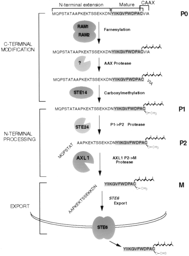
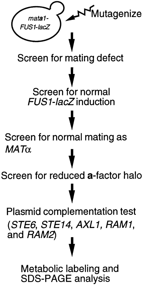
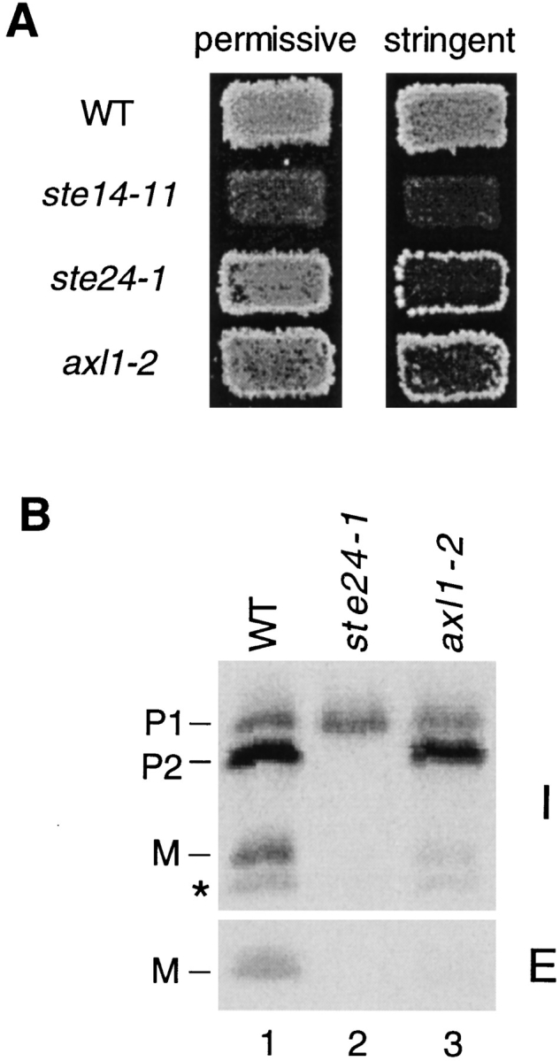
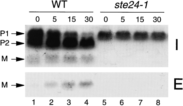
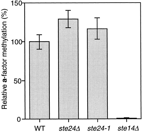
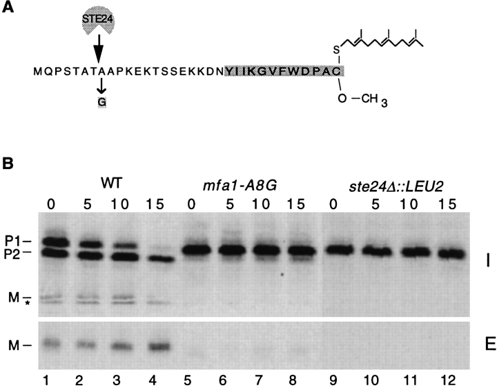
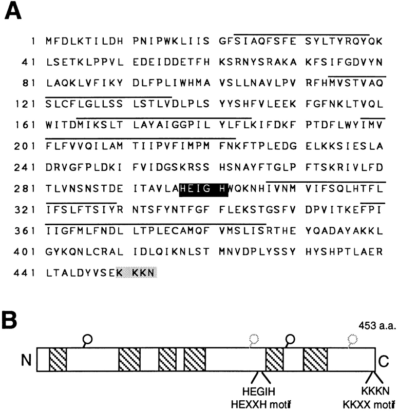
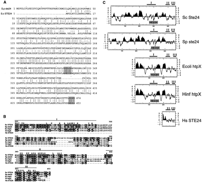
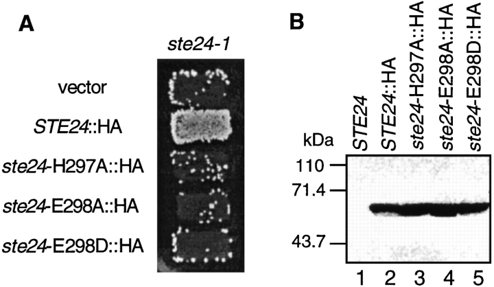
Many secreted bioactive signaling molecules, including the yeast mating pheromones a-factor and alpha-factor, are initially synthesized as precursors requiring multiple intracellular processing enzymes to generate their mature forms. To identify new gene products involved in the biogenesis of a-factor in Saccharomyces cerevisiae, we carried out a screen for MA Ta-specific, mating-defective mutants. We have identified a new mutant, ste24, in addition to previously known sterile mutants. During its biogenesis in a wild-type strain, the a-factor precursor undergoes a series of COOH-terminal CAAX modifications, two sequential NH2-terminal cleavage events, and export from the cell. Identification of the a-factor biosynthetic intermediate that accumulates in the ste24 mutant revealed that STE24 is required for the first NH2-terminal proteolytic processing event within the a-factor precursor, which takes place after COOH-terminal CAAX modification is complete. The STE24 gene product contains multiple predicted membrane spans, a zinc metalloprotease motif (HEXXH), and a COOH-terminal ER retrieval signal (KKXX). The HEXXH protease motif is critical for STE24 activity, since STE24 fails to function when conserved residues within this motif are mutated. The identification of Ste24p homologues in a diverse group of organisms, including Escherichia coli, Schizosaccharomyces pombe, Haemophilus influenzae, and Homo sapiens, indicates that Ste24p has been highly conserved throughout evolution. Ste24p and the proteins related to it define a new subfamily of proteins that are likely to function as intracellular, membrane-associated zinc metalloproteases.


Figures

Figure 1
Figure 1
Model for the pathway of a-factor biogenesis. The a-factor biosynthetic intermediates and the components of the a-factor biogenesis machinery are shown (see the text for more information). Several of the a-factor intermediates can be directly visualized by SDS-PAGE and are designated P0, P1, P2, and M (Chen et al., 1997). The a-factor precursor contains an NH2-terminal extension, a mature portion, and a COOH-terminal CAAX motif, as indicated at top. During a-factor biogenesis, the unmodified a-factor precursor (P0) undergoes COOH-terminal modification (prenylation, proteolytic cleavage of AAX, and carboxylmethylation) to yield the fully COOH-terminally modified species P1. Next, NH2-terminal proteolytic processing occurs in two distinct steps, the first (P1→ P2) cleavage removing seven residues from the NH2-terminal extension to yield the P2 species, and the second (P2→ M) cleavage generating mature a-factor, which is exported from the cell. Question marks indicate that the corresponding component has not yet been genetically identified.
Figure 2
Figure 2
Scheme of the screen for MAT a-specific sterile mutants. Details of this screen are described in the text.
Figure 7
Figure 7
Complementation analysis to identify the STE24 gene and map of the disruption construct used to generate the ste24Δ::LEU2 null allele. A map of the cloned DNA that complements ste24-1 is shown. Boxes containing arrows indicate the ORFs; the database designations are shown above their respective boxes. Shaded boxes below the map show subclones tested for complementation. The ability of DNA fragments to complement the mating- defective phenotype in strain SM3060 (ste24-1) is indicated at the right. Shown above the map is the γ-disruption vector used to construct the chromosomal ste24Δ::LEU2 allele. Restriction sites are as follows: Bg, BglII; HI, BamHI; Kp, KpnI; P, PstI; RV, EcoRV; Sa, SacI; Sc, ScaI; Sp, SphI; Xa, XbaI; Xh, XhoI.
Figure 3
Figure 3
Mating and a-factor biogenesis phenotypes of the axl1-2 and ste24-1 mutants. (A) Patch mating assay. Patches of the indicated MAT a strains were replica-plated onto a lawn of the MATα mating tester stain SM1068 on an SD plate containing 1% (permissive) or 0.05% (stringent) YEPD. Plates were incubated at 30°C for 2 d. Strains tested are wild-type (SM3039), ste14-11 (SM3064), ste24-1 (SM3044), and axl1-2 (SM3061); these three mutants were obtained in this study (see Materials and Methods). (B) Immunoprecipitation of a-factor. To examine their a-factor biogenesis profiles, wild-type and mutant cells were metabolically labeled for 5 min with [35S]cysteine and the label was chased for 15 min. Intracellular and extracellular fractions were separated, subjected to immunoprecipitation with a-factor antiserum, and the fate of a-factor was analyzed by SDS-PAGE, followed by autoradiography, as described in Materials and Methods. The intracellular precursor species (P1 and P2) and mature a-factor are indicated. The band below the mature M species marked with an asterisk (*) is an a-factor–related peptide (AFRP) that does not appear on all gels (Chen, 1993); AFRP will be described elsewhere (Chen, P., and S. Michaelis, manuscript in preparation). Strains shown are wild type (SM3104), ste24-1 (SM3105), and axl1-2 (SM3106).
Figure 4
Figure 4
Processing of the a-factor precursor in wild-type and ste24 mutant cells. Cells were pulse labeled with [35S]cysteine for 5 min and chased for the indicated times. Intracellular and extracellular fractions were separated and subjected to immunoprecipitation, electrophoresis, and autoradiography. Positions of the P1, P2, and M forms of a-factor are indicated. Strains are wild type (SM3104) and ste24-1 (SM3105).
Figure 5
Figure 5
Methylation of a-factor in wild-type and ste24 mutant cells. Cells were double-labeled with [35S]cysteine and S-adenosyl-l-[3H-methyl]methionine. Total cell extracts were prepared, immunoprecipitated with a-factor antiserum, and subjected to SDS-PAGE. The P1 species was excised from the gel. P1 was assayed for base-volatile [3H]methanol counts, reflecting its level of methylesterification and for [35S]cysteine incorporation to measure the total amount of labeled P1 that is present, as described in the Materials and Methods. The extent of P1 methylation in each strain was assessed by calculating the 3H cpm/35S cpm ratio. The level of P1 methylation in the mutant strains relative to wild type (100%) is indicated. Mean and standard error for three or four experiments is shown. Strains are wild type (SM3310), ste24-1 (SM3309), ste24Δ (SM3286), and ste14Δ (SM1871).
Figure 6
Figure 6
Comparison of a-factor processing in ste24 mutant cells and in cells bearing an mfa1 mutant plasmid defective in P1→ P2 cleavage. (A) Position of the mfa1-A8G mutation in a-factor. The mfa1-A8G mutation and the site of P1→ P2 cleavage are indicated. (B) Processing of a-factor in ste24 mutant cells and in a strain expressing the a-factor mutant mfa1-A8G. Cells were pulse labeled with [35S]cysteine, and the label was chased for the indicated times. Intracellular and extracellular fractions were separated, subjected to immunoprecipitation, electrophoresis, and phosphoimager analysis. Positions of the P1, P2, and M forms of a-factor are indicated. Strains are wild type (SM3310), ste24Δ (SM3286), and mfa1A8G (SM2059).
Figure 8
Figure 8
Ste24p predicted sequence and structure. (A) The amino acid sequence of Ste24p. Putative transmembrane segments predicted by hydropathy analysis (Kyte and Doolittle, 1982) are above the line. The zinc metalloprotease and di-lysine consensus motifs are shown in black and gray boxes, respectively. These sequence data are available from GenBank/EMBL/DDBJ under accession No. U77137. (B) Schematic structure of Ste24p. The hatched boxes show predicted transmembrane domains. The relative position of the zinc metalloprotease (HEXXH) and dilysine (KKXX) motifs are indicated. Circles indicate potential sites of N-glycosylation, based on the presence of an NXS/T consensus motif. Because the presence of a di-lysine retrieval signal suggests that the COOH terminus of Ste24p is cytosolically disposed, the sites rendered in light gray are predicted to be cytosolically oriented and thus are unlikely to be glycosylated.
Figure 9
Figure 9
Comparison of Ste24p with related proteins in other organisms. (A) The amino acid sequence of S. cerevisiae Ste24p (Sc Ste24) is compared with the S. pombe homologue (YAN5, protein accession number Q10071; here designated Sp ste24) using the Bestfit program (GCG software package). Identity is indicated by a line between the sequences and conserved changes are indicated by two dots (two corresponding bases in a codon). Gaps are shown as dots within the sequence. The zinc metalloprotease motif (HEXXH) and ER retrieval signal (KKXX) are shown in gray boxes. The Sc Ste24p and Sp ste24 proteins are 41.3% identical at the amino acid level. (B) Alignment of the COOH-terminal region of Ste24p and related sequences from other species. The COOH-terminal region of Ste24p homologues from five organisms are aligned by the Blosum-62 amino acid similarity matrix. Bars mark highly conserved subdomains (I, II, and III). Black boxes denote amino acid identity with S. cerevisiae Ste24p, and gray boxes denote amino acid similarity with Ste24p. Residues that are identical between more than two proteins in these subdomains are indicated above. The amino acid numbers of Sc Ste24p are shown. The accession numbers of E. coli htpX protein and H. influenzae htpX homologue protein are P23894 and P44840, respectively. The partial H. sapiens homologue (Hs STE24) sequence was assembled from the expressed sequence tag database sequences (Z43273, T35312, R54272, N76181, F11310, and T12172). (C) Comparison of hydropathy plots of Ste24p and related proteins. The algorithm of Kyte and Doolittle (1982) was used to generate hydropathy profiles for Ste24p homologues, with a window of 11 amino acids. Potential transmembrane regions are in black. The highly conserved subdomains shown in B are indicated above the boxes. The position of the zinc metalloprotease motif (HEXXH) is indicated.
Figure 10
Figure 10
Phenotype of ste24 HEXXH metalloprotease motif mutants. (A) Patch mating test of ste24 metalloprotease domain mutants. Patches of ste24-1 transformants harboring plasmids with the indicated HA epitope–tagged versions of wild-type and mutant STE24 were replica plate mated to a lawn of the MAT a mating tester strain SM1068 on an SD plate containing 0.05% YEPD. Strains are SM3069 (vector, pRS316), SM3071 (STE24::HA, pSM1107), SM3072 (ste24-H297A::HA, pSM1103), SM3073 (ste24E298A::HA, pSM1104), and SM3074 (ste24-E298D, pSM1105). (B) Steady-state levels of wild-type and mutant Ste24 proteins. Crude extracts prepared from strains bearing the indicated mutations were analyzed by immunoblotting with anti-HA 12CA5 antibodies. Extract from 0.5 OD600 U of cells was loaded in each lane. Lane 1, SM3082 (STE24 tagged in the reverse orientation, pSM1098); lane 2, SM3071 (STE24::HA, pSM1107); lane 3, SM3072 (ste24-H297A::HA, pSM1103); lane 4, SM3073 (ste24E298A::HA, pSM1104); and lane 5, SM3074 (ste24-E298D, pSM1105).

References

    1. Adames N, Blundell K, Ashby MN, Boone C. Role of yeast insulin-degrading enzyme homologues in propheromone processing and bud site selection. Science (Wash DC) 1995;270:464–467. - PubMed
    1. Altschul SF, Gish W, Miller W, Myers EW, Lipman DJ. Basic local alignment search tool. J Mol Biol. 1990;215:403–410. - PubMed
    1. Anderegg RJ, Betz R, Carr SA, Crabb JW, Duntze W. Structure of Saccharomyces cerevisiae mating hormone a-factor. Identification of S-farnesyl cysteine as a structural component. J Biol Chem. 1988;263:18236–18240. - PubMed
    1. Ashby MN, King DS, Rine J. Endoproteolytic processing of a farnesylated peptide in vitro. Proc Natl Acad Sci USA. 1992;89:4613–4617. - PMC - PubMed
    1. Ashby MN, Rine J. Ras and a-factor converting enzyme. Methods Enzymol. 1995;250:235–250. - PubMed

Publication types

MeSH terms

Associated data