. 1997 Jan 27;136(2):331-43.
doi: 10.1083/jcb.136.2.331.

Structure, subunit topology, and actin-binding activity of the Arp2/3 complex from Acanthamoeba

Affiliations

Structure, subunit topology, and actin-binding activity of the Arp2/3 complex from Acanthamoeba

R D Mullins et al. J Cell Biol. .

Abstract

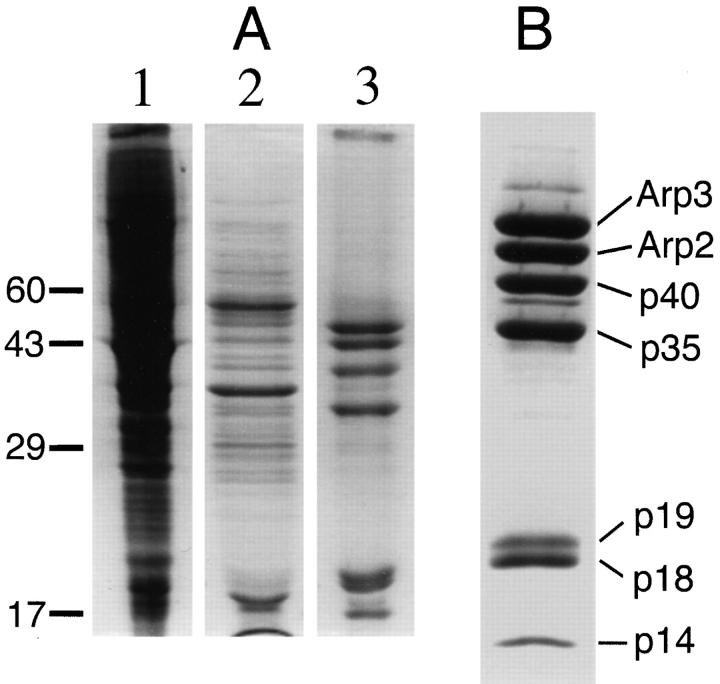
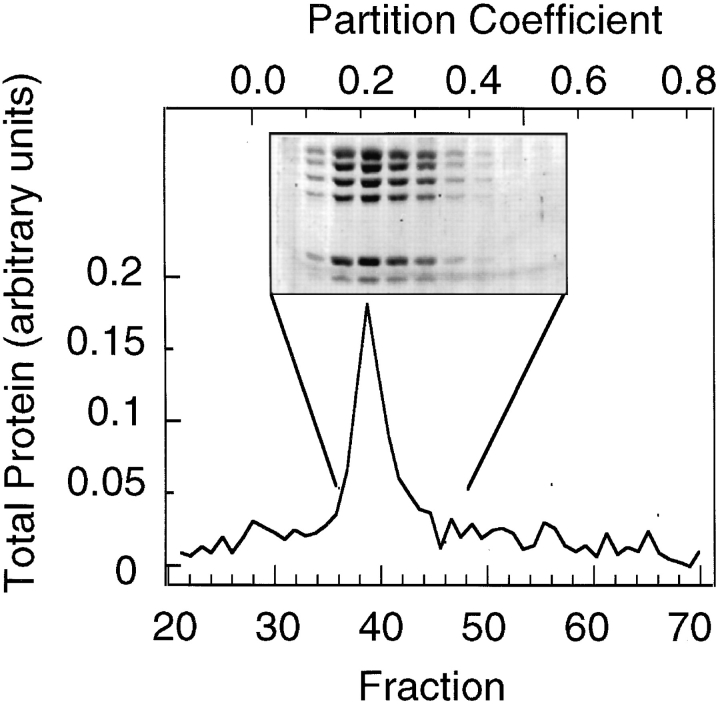
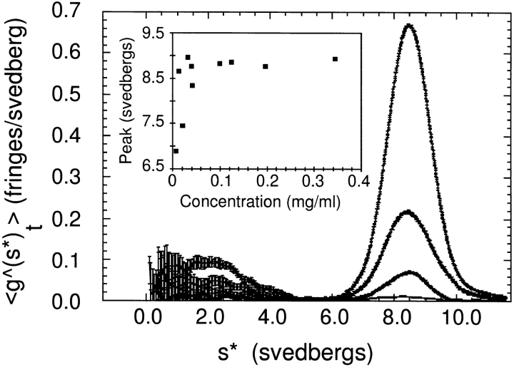
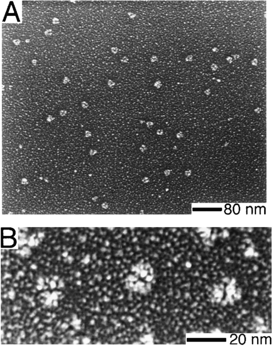
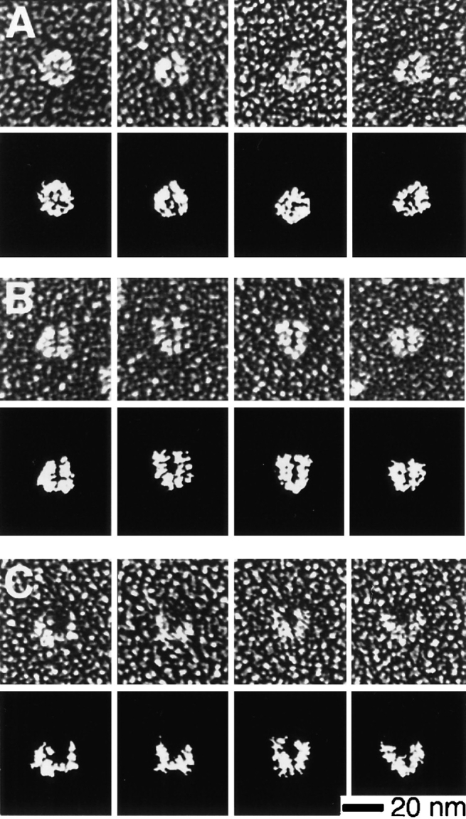
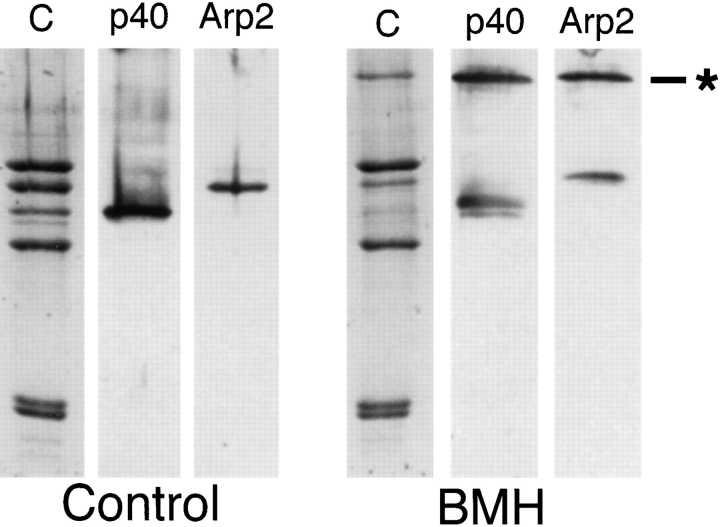
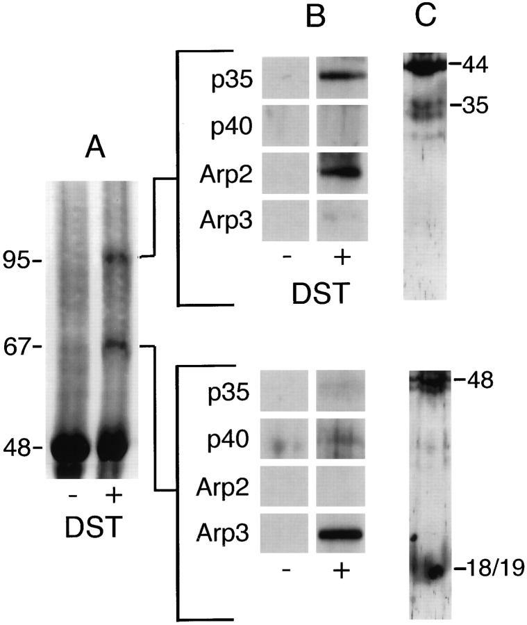
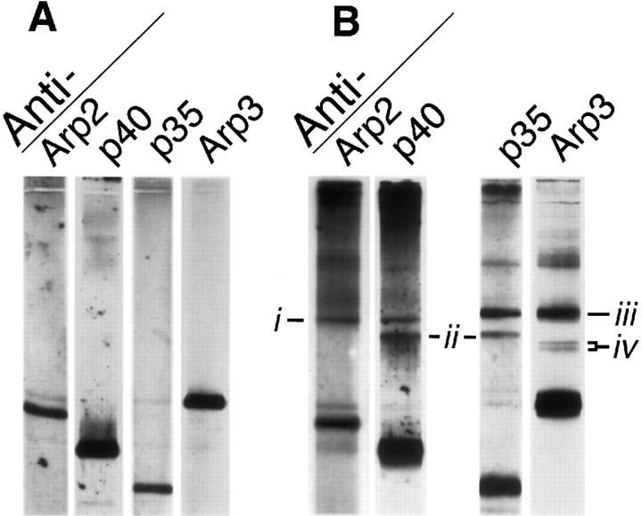
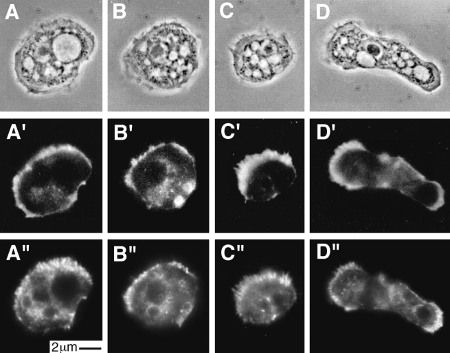
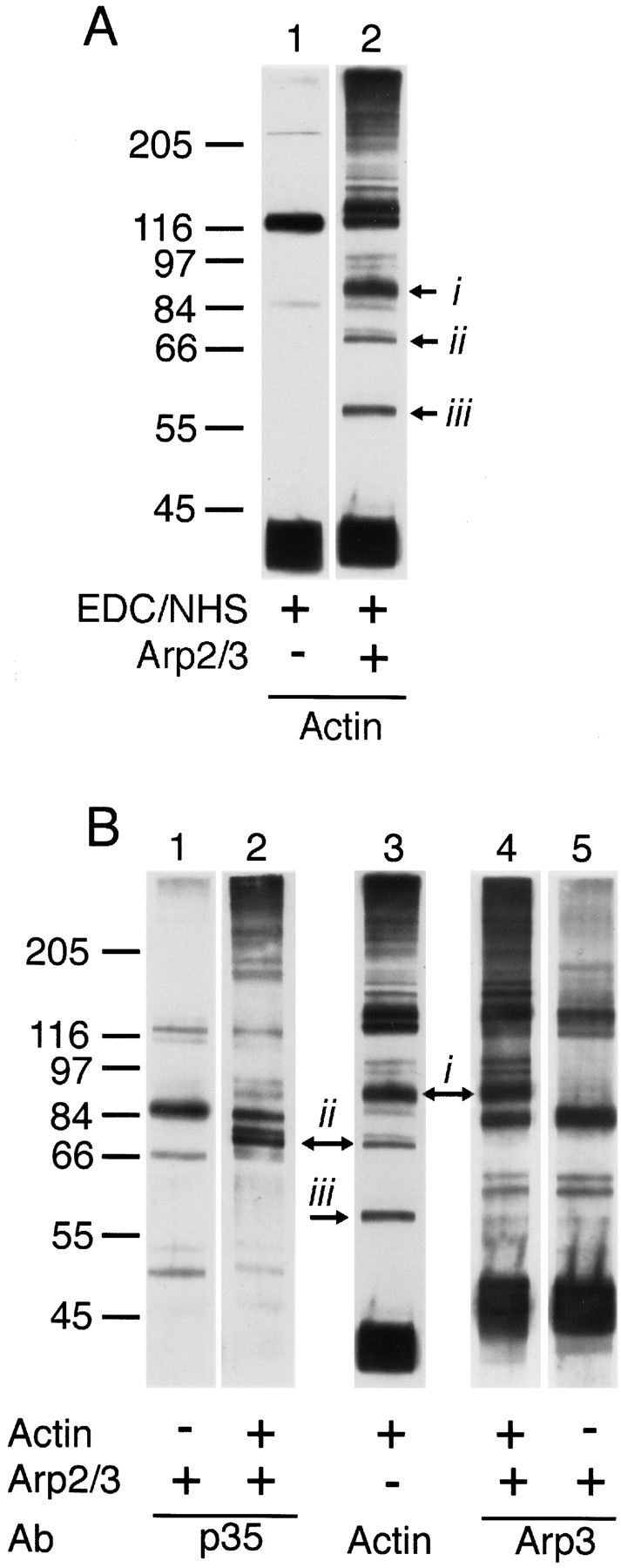
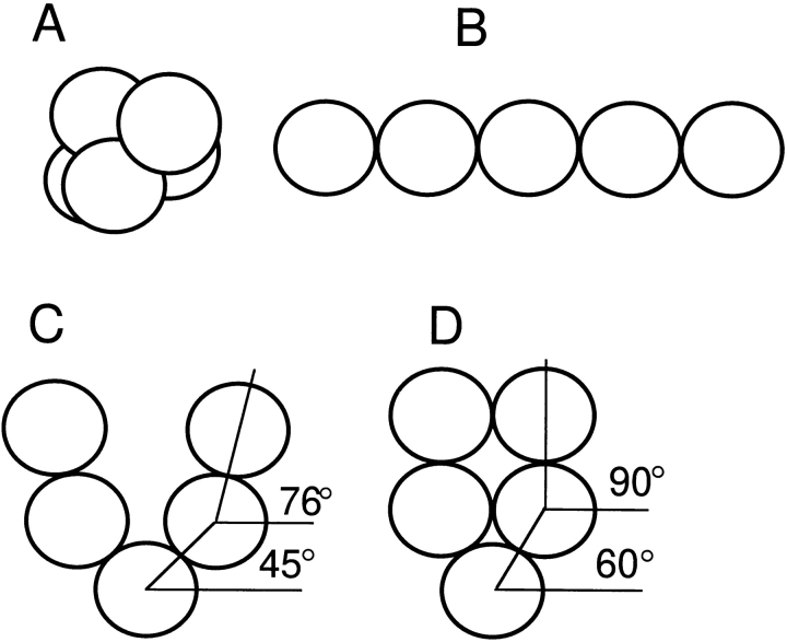
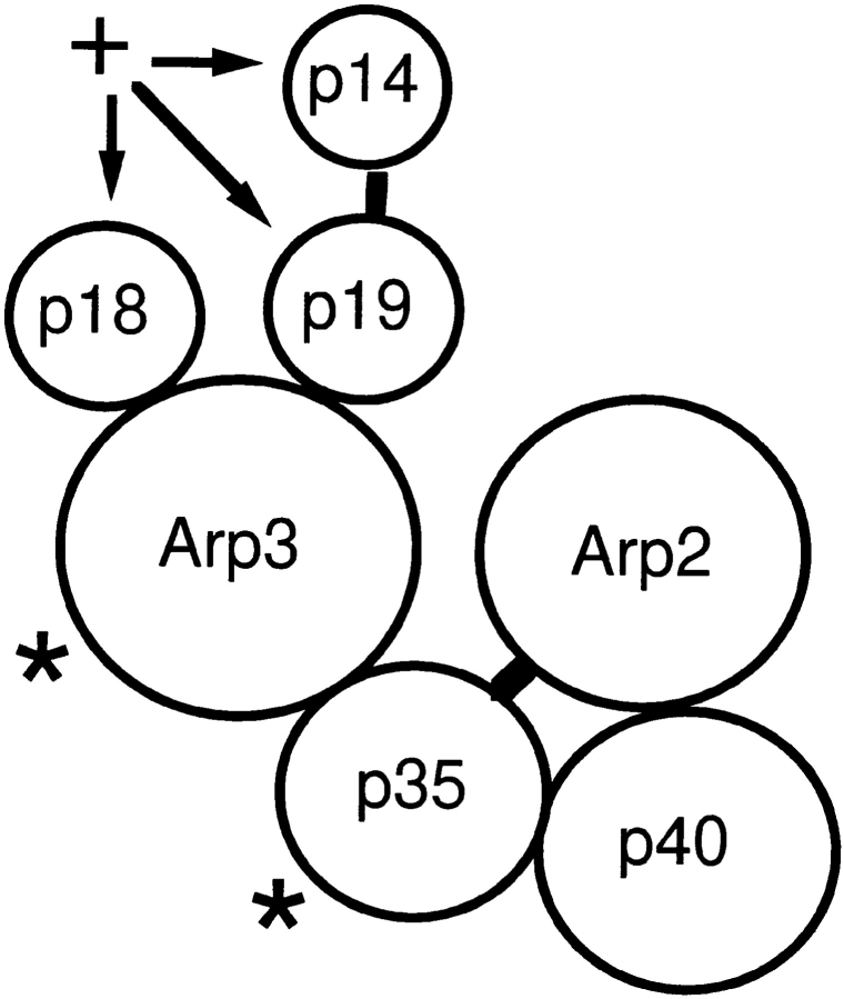
The Arp2/3 complex, first isolated from Acanthamoeba castellani by affinity chromatography on profilin, consists of seven polypeptides; two actin-related proteins, Arp2 and Arp3; and five apparently novel proteins, p40, p35, p19, p18, and p14 (Machesky et al., 1994). The complex is homogeneous by hydrodynamic criteria with a Stokes' radius of 5.3 nm by gel filtration, sedimentation coefficient of 8.7 S, and molecular mass of 197 kD by analytical ultracentrifugation. The stoichiometry of the subunits is 1:1:1:1:1:1:1, indicating the purified complex contains one copy each of seven polypeptides. In electron micrographs, the complex has a bilobed or horseshoe shape with outer dimensions of approximately 13 x 10 nm, and mathematical models of such a shape and size are consistent with the measured hydrodynamic properties. Chemical cross-linking with a battery of cross-linkers of different spacer arm lengths and chemical reactivities identify the following nearest neighbors within the complex: Arp2 and p40; Arp2 and p35; Arp3 and p35; Arp3 and either p18 or p19; and p19 and p14. By fluorescent antibody staining with anti-p40 and -p35, the complex is concentrated in the cortex of the ameba, especially in linear structures, possibly actin filament bundles, that lie perpendicular to the leading edge. Purified Arp2/3 complex binds actin filaments with a Kd of 2.3 microM and a stoichiometry of approximately one complex molecule per actin monomer. In electron micrographs of negatively stained samples, Arp2/3 complex decorates the sides of actin filaments. EDC/NHS cross-links actin to Arp3, p35, and a low molecular weight subunit, p19, p18, or p14. We propose structural and topological models for the Arp2/3 complex and suggest that affinity for actin filaments accounts for the localization of complex subunits to actin-rich regions of Acanthamoeba.


Figures

Figure 1
Figure 1
SDS-PAGE of fractions taken at each step in the purification of Arp2/3 complex from Acanthamoeba. Bands are stained with Coomassie blue. (A) An extract of soluble proteins (lane 1) was first passed over a DE-52 anion-exchange column in a low ionic strength buffer. Fractions that flowed through DEAE (lane 2) were pooled and loaded onto a poly-l-proline–Sepharose affinity column. Arp2/3 complex (lane 3) was eluted from the column with 0.4 M MgCl2. (B) Purified Arp2/3 complex, Arp3, Arp2, and five associated proteins.
Figure 2
Figure 2
Analytical gel filtration of Arp2/3 complex. All seven polypeptides of the purified Arp2/3 complex migrate as a single peak with a Stokes' radius of 5.3 nm. Conditions: 200 μl of purified Arp2/3 complex at 0.5 mg/ml was run on a 1 × 55-cm column of Sephacryl S300 equilibrated with 100 mM NaCl, 100 mM glycine, 0.2 mM MgCl2, 0.1 mM EGTA, 0.1 mM ATP, 1 mM DTT, 10 mM Tris-HCl, pH 7.0. Fractions of 0.4 ml were collected and protein concentration measured by the Bradford (1976) assay. Two column runs were averaged to produce the trace. Fractions from one run were precipitated with methanol/chloroform, and proteins were separated by SDS-PAGE and stained with Coomassie brilliant blue (inset).
Figure 3
Figure 3
Plots of the distribution of sedimentation coefficients of the Arp2/3 complex determined at various protein concentrations by sedimentation velocity ultracentrifugation. Conditions: The Arp2/3 complex was centrifuged in a Beckman model E centrifuge, AN-F Ti rotor at 40,000 rpm, 20°C, in buffer containing 100 mM NaCl, 100 mM glycine, 0.2 mM MgCl2, 0.1 mM EGTA, 0.1 mM ATP, 1 mM DTT, 10 mM Tris-HCl, pH 7.0. The samples were diluted by successive factors of three, giving a range of concentrations from 1.9 to 0.07 μM. (Inset) Plot of peak S value as a function of protein concentration. The sedimentation coefficient is stable over a wide range of concentrations but shifts to lower values below 70 nM.
Figure 4
Figure 4
Electron microscopy of purified Arp2/3 complex prepared by rotary replication. Conditions: Arp2/3 complex at 0.5 mg/ml in 150 mM NaCl, 0.2 mM MgCl2, 0.1 mM EGTA, 0.1 mM ATP, 1 mM DTT, 10 mM imidazole, pH 7.4, was mixed 1:1 with glycerol and sprayed onto freshly cleaved mica chips. Samples were vacuum-dried and rotary-replicated with platinum at 6° and carbon-coated at 60°. (A) Field of particles. (B) Higher magnification view of three molecules showing distinct bilobed structure.
Figure 5
Figure 5
Gallery of electron micrographs and camera lucida interpretations of purified Arp2/3 complex particles prepared by rotary replication. Conditions: See Fig. 4. (A) Approximately half (51%, n = 251) of the molecules surveyed displayed a compact, globular morphology with only a slightly bilobed appearance. (B) Nearly half of the molecules (44%) displayed a distinct bilobed morphology with a clearly defined central cleft. (C) A smaller number of molecules (5%) had a splayed, horseshoe shape with a large central cleft.
Figure 6
Figure 6
Chemical cross-linking of subunits in the Arp2/3 complex with the 1.61 nm, sulfhydryl-reactive cross-linker BMH. SDS-PAGE of reaction products. Conditions: Cross-linking was performed for 1 h at 24°C with 0.3 mg/ml purified Arp2/3 complex, 1.5 mM BMH, 10% DMSO, 150 mM NaCl, 0.2 mM MgCl2, 10 mM imidazole, pH 7.5. Reaction was quenched with an equal volume of 1 M Tris-HCl, pH 8.0. (Left) Control samples. (Right) BMH–cross-linked samples. C, Coomassie-stained polyacrylamide gel; p40, immunoblot probed with anti-p40; Arp2, immunoblot probed with anti-Arp2; *, cross-linked product. Arp2 and p40 are specifically cross-linked in high yield, forming a band of ∼85 kD recognized by both antibodies.
Figure 7
Figure 7
Chemical cross-linking of the Arp2/3 complex with the 0.64 nm, amine-reactive cross-linker DST. SDS-PAGE of reaction products. Conditions: Cross-linking was performed for 1 h at 24°C with 0.3 μg/ml purified Arp2/3 complex, 3 mM DST, 10% DMSO, 150 mM NaCl, 0.2 mM MgCl2, 10 mM imidazole, pH 7.5. Reaction was quenched with an equal volume of 1 M Tris-HCl, pH 8.0. (A) Coomassie-stained polyacrylamide gel. Lane 1, control; lane 2, DST–cross-linked samples. (B) Immunoblots of crosslinked bands probed with antibodies to Arp3, Arp2, p40, and p35. (C) Cross-linked bands were excised from wet gels, flash frozen, pulverized, and incubated overnight in 100 mM sodium periodate to oxidize DST cross-links. Uncross-linked polypeptides were separated by SDS-PAGE and stained with silver. Positions of the complex subunits from adjacent lanes on the same gel are marked. Arp2 is cross-linked to p35 and Arp3 to either p18 or p19.
Figure 8
Figure 8
Chemical cross-linking of the Arp2/3 complex (lane C) with the amine-reactive cross-linkers DSP and DSG. Conditions: Cross-linking was carried out for 1 h at 24°C with 0.14 mg/ml purified Arp2/3 complex, 1 mM DSP or DSG, 10% DMSO, 150 mM NaCl, 0.2 mM MgCl2, 0.1 mM EGTA, 10 mM imidazole, pH 7.5. Reaction was quenched with an equal volume of 1 M Tris-HCl, pH 8.0, and reaction products separated by SDS-PAGE and stained with Coomassie brilliant blue. The DSP–cross-linked band (*) was excised from a wet gel, flash-frozen, pulverized, and boiled in sample buffer containing β-mercaptoethanol to reduce the DSP cross-link. Uncross-linked polypeptides were separated by SDS-PAGE and stained with silver (Reduced). Positions of the complex subunits run in adjacent lanes on the same gel are marked. The 29-kD cross-linked product produced by DSP is a heterodimer of p19 and p14.
Figure 9
Figure 9
Chemical cross-linking of the Arp2/3 complex with the zero-length, heterobifunctional cross-linker EDC. Conditions: Cross-linking was carried out for 1 h at 24°C with 0.3 mg/ml purified Arp2/3 complex, 5 mM EDC, 5 mM NHS, 10% DMSO, 150 mM NaCl, 0.2 mM MgCl2, 0.1 mM EGTA, 10 mM imidazole, pH 7.5. Reaction was quenched with an equal volume of 1 M Tris-HCl, pH 8.0. Reaction products were analyzed by SDSPAGE and immunoblotting. Immunoblots were probed with monospecific antibodies against Arp3, Arp2, p40, and p35 at 1: 12,000 dilution. (A) Control complex. (B) Cross-linked complex. Cross-linking produces four new sets of bands: (i) a band of 85 kD recognized by antibodies against Arp2 and p40, (ii) a band of 75 kD recognized by antibodies against p40 and p35, (iii) a band of 88 kD recognized by antibodies against p35 and Arp3, and (iv) a doublet of bands at ∼66 and 70 kD recognized only by antibodies against Arp3.
Figure 10
Figure 10
Immunofluorescence localization of p35 and p40 in Acanthamoeba castellani. Cells fixed with 1% formaldehyde in 97.5% ice-cold methanol were stained with affinity-purified rabbit antibodies against either p35 (A and B) or p40 (C and D). (A–D) Phase contrast microscopy. (A″–D″) Localization of p35 and p40 detected with cy3-conjugated goat anti–rabbit secondary antibodies. (A′–D′) Actin filament localization with BODIPY-conjugated phalloidin. Both p35 and p40 were present throughout the cytoplasm and enriched in the cell cortex especially in fibrous structures within microspikes (C″) or entirely within the cell (A″). (E′and F′) Confocal images of 0.8 μm sections of fixed amebas stained with antibodies against p40 (E′ and F′). (E and F) Corresponding phase images. The confocal images emphasize the enrichment of p40 in regions of the cell cortex and the fibrous character of the staining pattern.
Figure 10
Figure 10
Immunofluorescence localization of p35 and p40 in Acanthamoeba castellani. Cells fixed with 1% formaldehyde in 97.5% ice-cold methanol were stained with affinity-purified rabbit antibodies against either p35 (A and B) or p40 (C and D). (A–D) Phase contrast microscopy. (A″–D″) Localization of p35 and p40 detected with cy3-conjugated goat anti–rabbit secondary antibodies. (A′–D′) Actin filament localization with BODIPY-conjugated phalloidin. Both p35 and p40 were present throughout the cytoplasm and enriched in the cell cortex especially in fibrous structures within microspikes (C″) or entirely within the cell (A″). (E′and F′) Confocal images of 0.8 μm sections of fixed amebas stained with antibodies against p40 (E′ and F′). (E and F) Corresponding phase images. The confocal images emphasize the enrichment of p40 in regions of the cell cortex and the fibrous character of the staining pattern.
Figure 11
Figure 11
Pelleting of Arp2/3 complex with actin filaments. Conditions: 50 mM KCl, 1 mM MgCl2, 1 mM EGTA, 10 mM imidazole, pH 7.0. A stock solution of 25 μM polymerized actin was diluted to various concentrations with 0.25 μM purified Arp2/3 complex. After 15 min at room temperature, we centrifuged the samples for 30 min at 24°C in a TLA 100 rotor at 200,000 g. Supernatants were analyzed by SDS-PAGE for depletion of complex subunits. Coomassie brilliant blue–stained gels were digitized with a Microtek ScanMaker III flat-bed scanner, and densities of gel bands were integrated by computer. Data shown are for depletion of p18+p19 and were fit to a rectangular hyperbola yielding an apparent K d of 2.3 ± 0.3 μM (R2 = 0.975).
Figure 12
Figure 12
Electron micrographs of single, negatively stained actin filaments in the absence (A) and presence (B) of Arp2/3 complex. Conditions: 20 μM actin was polymerized at 24°C for 30 min in 50 mM KCl, 1 mM MgCl2, 0.2 mM ATP, 1 mM EGTA, 10 mM imidazole, pH 7.0, and then diluted to 0.3 μM in the same buffer or buffer containing 2.3 μM Arp2/3 complex. Lightly glow-discharged, carboncoated grids were incubated on a drop of this solution and then stained with 1% uranyl formate according to the method of Aebi and Pollard (1987). The diameter of actin filaments decorated with Arp2/3 complex is 24 ± 3 nm (n = 15), compared to 7.5 ± 0.8 nm (n = 15) for actin filaments alone.
Figure 13
Figure 13
EDC cross-links actin to Arp3, to p35, and to one of the low molecular weight subunits of the Arp2/3 complex. Conditions: Cross-linking was carried out for 1 h at 24°C with 2 μM purified Arp2/3 complex, 4 μM filamentous actin, 5 mM EDC, 5 mM NHS, 10% DMSO, 50 mM KCl, 0.2 mM MgCl2, 0.1 mM EGTA, 10 mM imidazole, pH 7.5. Reaction was quenched with an equal volume of 1 M Tris-HCl, pH 8.0, and the products analyzed by SDS-PAGE and immunoblotting. Immunoblots were probed with monospecific antibodies against Arp3, p35, and actin at dilutions of 1:12,000, 1:6,700, and 1:10,000, respectively. Purified Arp2/3 complex and Acanthamoeba actin were incubated with EDC and NHS, and the reactions were quenched. (A) Immunoblots of EDC/NHS-cross-linked samples probed with monoclonal antiactin antibodies. Lane 1, EDC-treated actin filaments; lane 2, EDC-treated mixture of actin filaments and Arp2/3 complex showing three prominent new bands at (i) 97, (ii) 79, and (iii) 59 kD. (B) Immunoblots of EDC/NHS-cross-linked samples probed with antibodies against p35 (lanes 1 and 2), actin (lane 3), or Arp3 (lanes 4 and 5). The cross-linking reactions contained Arp2/3 complex, actin filaments, or both as indicated with +. Arrows denote new bands recognized by the anticomplex antibodies that correspond to new bands recognized by antiactin antibodies.
Figure 14
Figure 14
Hydrodynamic bead models of the Arp2/3 complex. Models assume five rigidly linked, identical 4.52-nm-diam spheres, each with a partial specific volume of 0.74 cm3/gm. Each model, thus, has a molecular mass of 197 kD. The partial specific volume and hydration (0.323 g/g) used in all calculations were based on amino acid analysis. Hydrodynamic parameters for each configuration were calculated by the method of Riseman and Kirkwood (1956). (A) Globular, double-tetrahedral arrangement, 10.3 S. (B) Linear model, 8.1 S. (C) Open horseshoe-shaped model, 8.7 S. (D) Compact horseshoe-shaped model, 9.5 S. The best agreement with experimental data was given by model C.
Figure 15
Figure 15
Chemical crosslinking model of the Arp2/3 complex. Lines represent chemical cross-links made between subunits with various cross-linkers. Line lengths are proportional to cross-linker spacer arms and subunit sizes are proportional to apparent molecular weights. Both subunits denoted with a * and one of the three subunits denoted with a + can be cross-linked to actin by EDC/NHS.

Similar articles

Cited by

References

    1. Ackers GK. A new calibration procedure for gel filtration columns. J Biol Chem. 1967;242:3237–3238.
    1. Aebi U, Pollard TD. A glow discharge unit to render electron microscope grids and other surfaces hydrophilic. J Electron Micro Tech. 1987;7:29–33. - PubMed
    1. Aebi U, Fowler WE, Isenberg G, Pollard TD, Smith PR. Crystalline actin sheets: their structure and polymorphism. J Cell Biol. 1981;91:340–351. - PMC - PubMed
    1. Balasubramanian MK, Feoktistova A, McCollum D, Gould K. Fission yeast Sop2p: a novel and evolutionarily conserved protein that interacts with Arp3p and modulates profilin function. EMBO (Eur Mol Biol Organ) J. 1996;15:6426–6437. - PMC - PubMed
    1. Bradford M. A rapid and sensitive method for the quantitation of microgram quantities of protein utilizing the principle of protein dye binding. Anal Biochem. 1976;72:248–254. - PubMed

Publication types

MeSH terms